
Physiology
Muscle Under the Microscope - Episode 8: Muscle Injuries and the Repair Process
September 6, 2026
In the previous episodes we moved from the microscopic architecture of the muscle fiber to actin, myosin, the neuromuscular junction, calcium and hypertrophy. Now we reach the more difficult part of the story: what happens when muscle tissue is injured? A muscle injury does not simply mean that a fiber has torn. It means that an entire cellular and molecular ecosystem enters a coordinated sequence of degeneration, inflammation, repair and remodeling.
Skeletal muscle has a remarkable capacity to regenerate after many acute injuries. The process depends on satellite cells, immune cells, the extracellular matrix, blood vessels and local signaling. At the same time, prolonged inflammation, repeated injury or a poorly controlled environment can favor fibrosis and loss of function. Repair is therefore not the simple reversal of a tear, but a carefully regulated reconstruction.
1. What is a muscle injury?
A muscle injury is a disruption of the integrity and function of muscle tissue. It can range from microscopic changes and membrane damage to major structural disruption of fibers and fascicles. The type, location and severity of the injury strongly influence how the tissue will repair itself.
In sport, muscles may experience contusions, strains, damage associated with eccentric contractions and tears. Not every post-training ache represents a major structural injury, and delayed-onset muscle soreness should not automatically be equated with a tear. Biologically, the extent of tissue disruption is more important than the subjective intensity of pain.
2. The first event - fibers and membranes are damaged
After injury, parts of muscle fibers can lose the integrity of the sarcolemma and internal structures. Myofibrils may be damaged, sarcomere organization may be disturbed, and molecules released into the extracellular environment can signal that tissue has been injured.
In more significant injuries, segments of the fiber may undergo necrosis, with local bleeding and accumulation of cellular debris. Damaged tissue cannot simply be glued back together. It must be cleared, and regenerative cell populations must then be activated in an appropriate sequence.
3. Inflammation - why the first response is not the muscle's enemy
Inflammation begins rapidly after injury and is one of the first stages of repair. Immune cells are recruited to the damaged area, where they help remove destroyed tissue and release signals that alter the local environment.
Macrophages are particularly important. Early in repair they contribute to debris clearance and the inflammatory response, while later their functional state changes in ways that support resolution of inflammation, muscle-cell differentiation and tissue remodeling. Controlled acute inflammation supports repair; the problem arises when the response becomes prolonged or dysregulated.
4. Satellite cells - the regenerative reserve of muscle
Satellite cells, also called muscle stem cells, normally remain quiescent between the sarcolemma and basal lamina. When an appropriate injury signal appears, they leave quiescence and enter a program of proliferation and differentiation.
These cells are not simply passive reserves. They form a heterogeneous population, and some activated cells become myogenic progenitors that contribute to repair, while others replenish the satellite-cell pool. In this way, muscle attempts to repair the injury without exhausting its regenerative source.
5. From satellite cell to myoblast
After activation, satellite cells proliferate and generate muscle progenitor cells called myoblasts. These cells align, differentiate and can fuse with one another or with existing muscle fibers to contribute to restoration of fiber structure.
The process is regulated by myogenic regulatory factors including MyoD, Myf5 and myogenin. Their expression changes across activation and differentiation. Ultimately, newly incorporated cells can contribute to functional muscle regions and restoration of the contractile apparatus.
6. Clearing the area - why debris must be removed
Regeneration cannot proceed efficiently in an environment filled with necrotic debris. Macrophages and other phagocytic cells remove cellular fragments and help shift the inflammatory environment. This prepares the tissue for reconstruction.
Clearance is not simply mechanical. Immune cells send signals to satellite cells and other local populations, while the extracellular matrix and vascular environment are changing in parallel. Repair is therefore a conversation between several types of cells.
7. The extracellular matrix - the invisible scaffold of reconstruction
Around muscle fibers lies an extracellular matrix made of collagen, laminin, fibronectin and other components. It is not merely filler. The matrix provides mechanical support and influences satellite-cell behavior, cell migration and tissue organization during repair.
During normal regeneration, the matrix is remodeled to support reconstruction. When collagen deposition becomes excessive and persistent, fibrosis can develop. Fibrotic tissue can restore mechanical continuity, but it does not reproduce the contractile properties and architecture of healthy muscle.
8. Formation of new fibers and their maturation
As myoblasts differentiate and fuse, immature muscle structures appear. Newly regenerated fibers initially differ from mature fibers and pass through a maturation stage in which sarcomeres become organized, extracellular relationships are restored, and vascular and metabolic support adapt to the new tissue.
Regeneration is not just the production of a new membrane. For a fiber to become functional, relationships with the nervous system, blood vessels, extracellular matrix and neighboring tissues must also be restored. Biological recovery can therefore continue long after pain has decreased.
9. Blood vessels and oxygen supply
A muscle region undergoing repair needs nutrients, oxygen and removal of metabolic products. Vascular cells and angiogenic signals participate in restoring the capillary network within injured tissue.
Vascular support also matters because muscle is a highly active metabolic tissue. As regenerated fibers mature, the vascular environment must be capable of supporting the new structure. Muscle repair is therefore linked to regeneration of several compartments at the same time.
10. The nerve has to return to the story
A structurally repaired muscle is not fully functional if it cannot be activated properly. Recovery includes restoration of neuromuscular relationships and reorganization of motor control. Depending on the type and severity of injury, reinnervation and recovery of coordination can follow a different timeline from muscle tissue healing itself.
This explains why an athlete may feel that the muscle is «healed» because pain has disappeared, yet still be unable to produce the same force or coordination. Recovery of function is more than disappearance of the symptom.
11. Fibrosis - when repair leaves too much scar tissue
After injury, extracellular-matrix deposition is necessary to stabilize and reconstruct the tissue. But when this response becomes excessive or inflammation persists, too much collagen can accumulate and fibrosis can develop.
Fibrosis can reduce tissue elasticity, alter force transmission and create a microenvironment that is unfavorable for muscle regeneration. In repeated or chronic injury, the interaction among inflammation, stromal cells and extracellular matrix can shift the balance from regeneration toward scarring.
12. Why repeated injuries can be a bigger problem
A well-resolved acute injury gives muscle an opportunity to move through an orderly sequence of inflammation, regeneration and remodeling. When new trauma occurs before tissue has fully regained function, repair mechanisms can be disrupted.
Repeated trauma can maintain inflammation, increase matrix deposition and compromise regenerative quality. This is one reason return to loading should consider not only pain, but also strength, motor control, mobility and progressive tolerance to load.
13. Muscle injury in Strongman - what happens under extreme loads
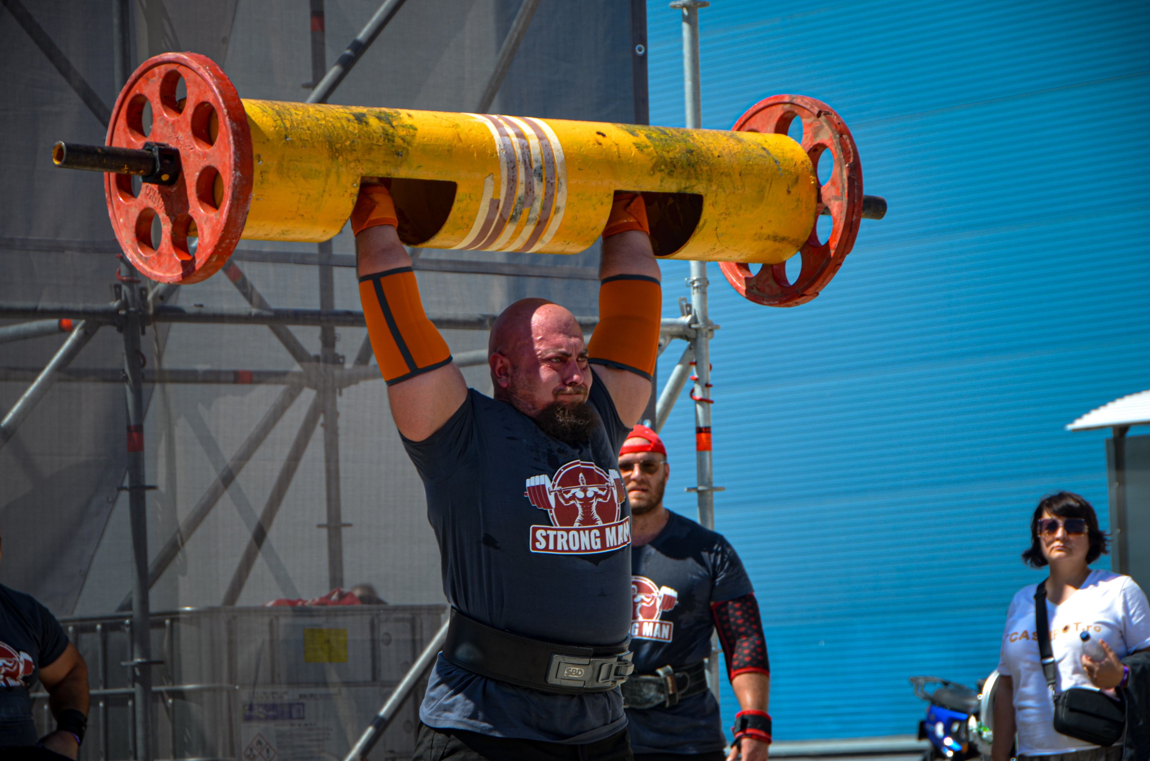
Strongman exposes muscles to unusual combinations of tension, joint positions, high loads and fatigue. Heavy deadlifts, Atlas stones, loaded carries, log press and other events can challenge muscle fibers, tendons, fascia and passive tissues at the same time.
When injury occurs, return is not simply a matter of waiting for inflammation to disappear. Tissue must regain the ability to tolerate tension and relearn how to produce force in the context of a specific event. Progressive rehabilitation helps rebuild this capacity without prematurely overloading tissue that is still vulnerable.
14. Recovery - why time alone is not always enough
Biological healing has its own timeline, but function recovers through the interaction between spontaneous healing and appropriately graded loading. In practice, return to sport depends on injury severity, location, tissue status, symptoms and response to loading.
Modern rehabilitation aims to preserve or regain function within tolerated limits and then progressively increase mechanical demands. The final goal is not merely absence of pain, but the ability to tolerate again the speed, force and fatigue specific to the sport. For a Strongman athlete, this may mean progressing from controlled movements to heavy loading and then to event-specific work.
15. Conclusion - muscle does not simply heal, it rebuilds itself
After muscle injury, tissue passes through a complex sequence: damage, inflammation, debris clearance, satellite-cell activation, progenitor proliferation and differentiation, fiber fusion and maturation, extracellular-matrix remodeling, vascular restoration and recovery of neuromuscular control. Each stage prepares the next.
The most important point is that repair does not automatically mean perfect restoration of the original structure. Sometimes tissue regenerates extremely well; at other times fibrosis or reduced function remains, especially after severe or repeated injury. For an athlete, the real goal is recovery of a structure and function robust enough for the specific demands of sport.
Under the microscope, muscle recovery is a story of cooperation: satellite cells, macrophages, blood vessels, extracellular matrix and muscle fibers work together. When this reconstruction is accompanied by a progressive return to loading, tissue does more than survive the injury. It attempts to become capable of producing force again.
Scientific bibliography
1. Tidball JG. Inflammatory processes in muscle injury and repair. American Journal of Physiology-Regulatory, Integrative and Comparative Physiology. 2005;288:R345-R353.
2. Charge SBP, Rudnicki MA. Cellular and molecular regulation of muscle regeneration. Physiological Reviews. 2004;84(1):209-238. DOI: 10.1152/physrev.00019.2003.
3. Yin H, Price F, Rudnicki MA. Satellite cells and the muscle stem cell niche. Physiological Reviews. 2013;93(1):23-67. DOI: 10.1152/physrev.00043.2011.
4. Murphy MM, Keefe AC, Lawson JA, Olson EN, Kardon G. Regeneration and fibrosis in skeletal muscle. Nature Reviews Molecular Cell Biology. 2011;12:161-174.
5. Relaix F, Zammit PS. Satellite cells are essential for skeletal muscle regeneration: the cell on the edge returns centre stage. Development. 2012;139(16):2845-2856.
6. Tidball JG, Villalta SA. Regulatory interactions between muscle and the immune system during muscle regeneration. American Journal of Physiology-Regulatory, Integrative and Comparative Physiology. 2010;298:R1173-R1187.
7. Saclier M, Yacoub-Youssef H, Mackey AL, et al. Differentially activated macrophages orchestrate myogenic precursor cell fate during human skeletal muscle regeneration. Stem Cells. 2013;31(2):384-396.
8. Dumont NA, Bentzinger CF, Sincennes MC, Rudnicki MA. Satellite cells and skeletal muscle regeneration. Comprehensive Physiology. 2015;5(3):1027-1059.
9. Fry CS, Lee JD, Jackson JR, Kirby TJ, Stasko SA, Liu H, Dupont-Versteegden EE, McCarthy JJ, Peterson CA. Regulation of the muscle fiber microenvironment by activated satellite cells during myogenesis. Cell. 2014;158(2):288-299.
10. Jiang H, et al. MuSCs and IPCs: roles in skeletal muscle homeostasis, aging and injury. Cell and Molecular Life Sciences. 2024;81:67. DOI: 10.1007/s00018-023-05096-w.
11. Crosbie R, et al. Role of microenvironment on muscle stem cell function in health, adaptation, and disease. International Review of Cell and Molecular Biology. 2024. DOI: 10.1016/bs.ctdb.2024.02.002.
12. Beyond the bulk: overview and novel insights into the dynamics of muscle satellite cells during muscle regeneration. 2024. PMID: 39327631.
More From The Journal

Training
The Science of Strongman Training - Episode X: Competition Programming
How to build a complete Strongman competition preparation block: calendar, events, volume, intensity, specificity, simulations, autoregulation, tapering and fatigue management.
Training
The Science of Strongman Training - Episode IX: Peaking
Peaking in Strongman: how to turn months of training into maximal competition performance by reducing fatigue, preserving adaptations and managing specificity.