
Physiology
Muscle Under the Microscope - Episode 6: Calcium and Muscle Contraction
September 6, 2026
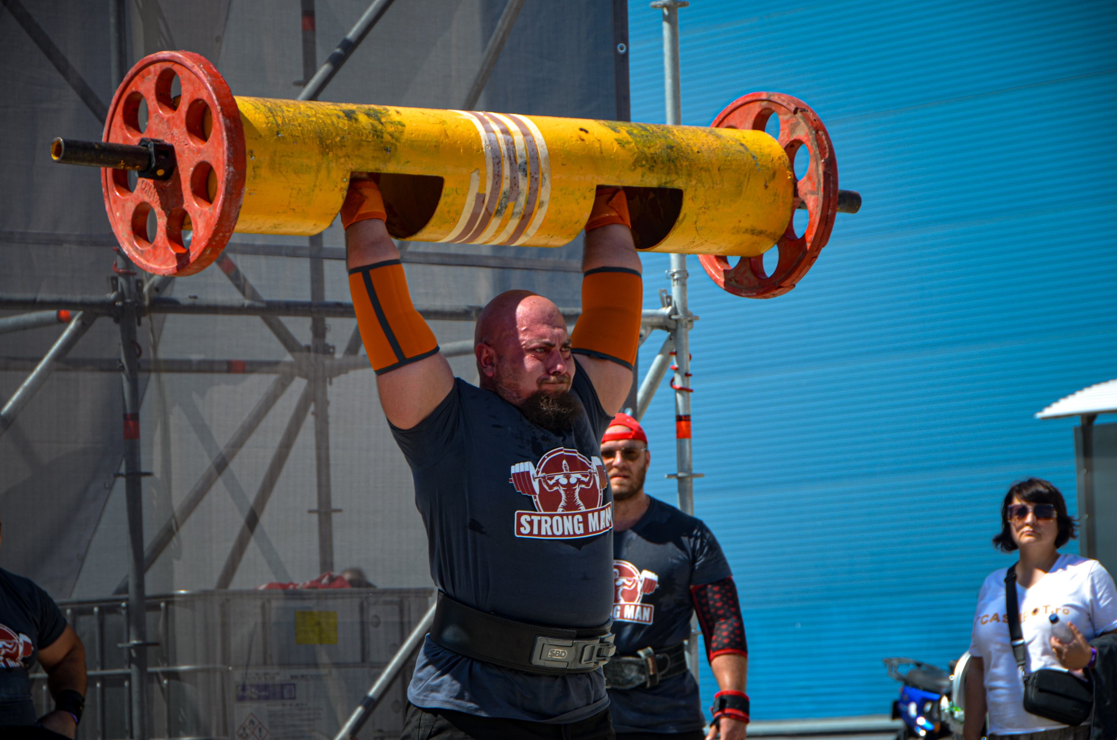
After following the microscopic architecture of muscle, fiber types, fast versus slow fibers, actin and myosin, and the neuromuscular junction in the previous episodes, we now reach one of the most important molecules in the story of contraction: calcium, Ca2+. Calcium is not simply a mineral in the body. In skeletal muscle fibers, rapid changes in free cytoplasmic Ca2+ are the molecular signal that permits the transition from rest to activation of the contractile apparatus.
This episode follows calcium from the sarcoplasmic reticulum to troponin and the actin-myosin filaments. We will examine how Ca2+ is released through RyR1, how its effect is terminated by SERCA-dependent reuptake, why the amplitude and duration of the calcium signal matter for force, and how these mechanisms connect to heavy Strongman efforts.
1. Why calcium is essential for muscle contraction
A skeletal muscle fiber can be viewed as a system in which an electrical signal must be converted extremely quickly into a chemical event and then into mechanical force. This process is known as excitation-contraction coupling. The action potential does not directly pull on actin and myosin. Instead, it triggers Ca2+ release from the sarcoplasmic reticulum, and the resulting transient rise in free myoplasmic Ca2+ activates the contractile apparatus.
At rest, free cytoplasmic Ca2+ is kept very low compared with the total amount of calcium stored in the sarcoplasmic reticulum. This difference creates a powerful signal: when release channels open, Ca2+ rapidly enters the myoplasmic space and changes the state of the troponin-tropomyosin regulatory system.
2. Where is calcium stored in the muscle fiber?
The main rapid Ca2+ reservoir for skeletal muscle contraction is the sarcoplasmic reticulum, a specialized membrane network surrounding the myofibrils. Its terminal regions lie very close to the transverse tubules, forming specialized structures called triads.
Within the reticulum, calcium is buffered and stored with proteins such as calsequestrin, allowing a large amount of Ca2+ to be available without all of it remaining free. Spatial organization is crucial. The reticulum is not simply a diffuse reservoir; it is part of a microscopic system designed for rapid release and reuptake of calcium.
3. From the action potential to Ca2+ release
After the neuromuscular junction transmits the signal, the muscle fiber generates its own action potential. It propagates along the sarcolemma and enters the depth of the fiber through the transverse tubular system. This brings the electrical signal close to numerous contractile units.
CaV1.1, also called the dihydropyridine receptor, is located in the membrane of the T-tubules. In skeletal muscle, its major role in excitation-contraction coupling is as a voltage sensor. Changes in membrane potential produce a conformational change that is functionally transmitted to the calcium-release machinery of the sarcoplasmic reticulum.
The central partner in the reticulum is RyR1, the type 1 ryanodine receptor. Functional communication between CaV1.1 and RyR1 permits rapid opening of RyR1 channels and massive but transient release of Ca2+ into the myoplasm. This is one of the fundamental steps of excitation-contraction coupling.
4. RyR1 - the gate through which calcium leaves the reticulum
RyR1 is a large Ca2+ release channel located in the membrane of the sarcoplasmic reticulum. During muscle activation, RyR1 channels open in a highly coordinated manner, allowing a rapid rise in free Ca2+ near the myofilaments.
The importance of RyR1 extends beyond the idea of a simple pore. The channel is integrated into a large macromolecular complex containing proteins that contribute to organization, stability, and regulation of calcium release. This architecture helps muscle fibers generate fast, repeatable signals that are essential for repeated contractions.
5. Calcium meets troponin
After release, Ca2+ diffuses through the myoplasm and reaches the thin filaments. One of its key targets is troponin, a regulatory protein complex associated with actin and tropomyosin. In skeletal muscle, Ca2+ binds primarily to troponin C.
Binding of Ca2+ to troponin C changes the relationships and positioning of the troponin-tropomyosin complex. Tropomyosin shifts on the actin filament, exposing more of the sites through which myosin heads can interact with actin.
An important nuance is that calcium does not directly pull the filaments and is not a mechanical motor. It acts as a regulatory signal that permits the actomyosin apparatus to enter the active state. The mechanical motor is the actin-myosin cross-bridge cycle, powered by ATP.
6. How does Ca2+ turn a signal into force?
Once sites on actin become accessible, myosin heads can enter the cross-bridge cycle. Myosin binds actin, changes conformation, contributes to filament sliding, and then detaches so a new cycle can begin. ATP is essential both to drive the mechanical cycle and to release myosin from actin.
The amount of free Ca2+ and the duration of its signal influence how strongly the contractile apparatus is activated. In simplified terms, a sufficiently strong signal activates more regions of the thin filament and increases the probability of active cross-bridge formation. The real relationship between Ca2+, activation, and force is dynamic and depends on fiber state, length, velocity, temperature, and recent activation history.
7. Why does one nerve discharge produce a short contraction?
A single action potential is very brief, but it can trigger a Ca2+ transient sufficient to activate contraction. After the Ca2+ peak, free Ca2+ begins to fall, and the systems responsible for relaxation regain control.
Mechanical contraction is not a perfect copy of the electrical signal. Calcium rises and falls quickly, while cross-bridge cycling and tension development have their own time courses. Therefore, one action potential produces a twitch, while repeated stimulation can overlap contractions and generate greater sustained tension.
8. SERCA - the pump that returns calcium to the reticulum
For the fiber to relax, Ca2+ cannot remain free in the cytoplasm at levels associated with activation. A major part of relaxation is active reuptake of Ca2+ into the sarcoplasmic reticulum by SERCA pumps, Ca2+-transporting ATPases that use energy from ATP.
SERCA is essential for repeated contraction. It does not produce force in the actomyosin sense, but without calcium reuptake the fiber could not efficiently return toward rest or handle a normal succession of contractions.
This creates a fundamental pair: RyR1 releases Ca2+ for activation, while SERCA retrieves it for relaxation and restoration of the intracellular calcium reservoir. Contraction and relaxation are therefore two phases of the same calcium-handling circuit.
9. Calcium and impulse frequency
When a second action potential arrives before the effect of the first Ca2+ transient has completely disappeared, activation can overlap. In a repeatedly stimulated fiber, calcium concentration and contractile activation reflect not only one impulse but the recent history of stimulation.
This temporal summation helps explain the transition from twitch to tetanus. In strength activity, the nervous system controls discharge frequency and motor-unit recruitment to regulate tension. Calcium is the microscopic link between stimulation frequency and the active state of the contractile apparatus.
10. Calcium does not automatically mean maximal force
It is tempting to say that more Ca2+ simply means more force. Reality is more complex. Force depends on activation, but also on motor-unit recruitment, discharge frequency, sarcomere length, fiber type, muscle architecture, myosin properties, joint mechanics, tendon behavior, and technique.
The amount of Ca2+ released also varies with physiological conditions. Repeated activity, fatigue, and local changes in the intracellular environment can influence calcium release and contractile responsiveness. Thus, Ca2+ is a central activation factor, not a single explanation for strength performance.
11. What happens to calcium during muscle fatigue?
During muscle fatigue, calcium-related mechanisms can become part of the overall decline in force. Depending on exercise type and experimental conditions, changes may occur in Ca2+ release, the sensitivity of the contractile apparatus to Ca2+, and the ability to re-sequester calcium.
It is important to avoid the idea that fatigue simply means that the muscle has «run out of calcium». The sarcoplasmic reticulum has storage, release, and reuptake systems, while fatigue is a multifactorial phenomenon involving central and peripheral changes, metabolic disturbances, and alterations at several links in the force-production chain.
12. Calcium and fast versus slow fibers
Muscle fibers do not all have identical calcium-handling kinetics. Differences between fiber types include the properties of proteins and systems involved in calcium release and reuptake. Fast fibers are adapted for rapid activation and relaxation, whereas slow fibers are specialized for sustaining activity.
SERCA isoforms and other proteins involved in Ca2+ homeostasis contribute to these differences. Consequently, the same neural command can have a different time course in fibers with distinct contractile properties.
13. Calcium during Strongman efforts
In a maximal deadlift, a heavy log press, or an Atlas stone lift, the external force we see is the final result of a very long chain. The brain generates the command, motor neurons discharge, the neuromuscular junction transmits the signal, the muscle fiber produces an action potential, CaV1.1 and RyR1 trigger Ca2+ release, troponin permits filament activation, and actin and myosin turn that activation into tension.
For maximal strength events, timing is critical. It is not enough for the muscle to be large. The system must rapidly activate many motor units and convert that activation into mechanical force. Calcium sits at the center of every active fiber, exactly where the electrical command meets the contractile machinery.
14. What happens when the calcium system is disturbed?
Because the mechanism is so integrated, abnormalities in proteins involved in excitation-contraction coupling can have major consequences. Mutations affecting RyR1 or other components of the calcium-release machinery are associated with muscle diseases and impaired muscle function.
These disorders show how precisely the system must be regulated. Insufficient effective release can impair contraction, while dysregulated Ca2+ channel control can produce pathological effects. In healthy muscle, the goal is not a permanently high Ca2+ concentration, but a rapid, localized and transient rise followed by restoration of the resting state.
15. Conclusion - calcium is the signal that turns the motor on
Calcium occupies a unique position in skeletal muscle physiology. It is the link connecting electrical excitation to mechanical activation. The signal starts with the action potential, travels through the T-tubule system to CaV1.1, reaches RyR1 functionally, releases Ca2+ from the sarcoplasmic reticulum, and activates troponin. From there, actin and myosin can enter the cross-bridge cycle and generate tension.
The shutdown phase is equally important. SERCA retrieves Ca2+, free calcium falls, troponin and tropomyosin move back toward the resting state, and the fiber becomes ready for another impulse. Muscle contraction is therefore a continuous dialogue between electricity, calcium, and mechanics.
For Strongman, every kilogram lifted is the final expression of this microscopic process. Behind an Atlas stone, a heavily loaded barbell, or a very heavy log press are billions of coordinated molecular events. At the center of every activated muscle fiber is a small Ca2+ wave telling the contractile apparatus that it is time to work.
Scientific bibliography
1. Shishmarev D. Excitation-contraction coupling in skeletal muscle: recent progress and unanswered questions. Biophysical Reviews. 2020;12:143-153. DOI: 10.1007/s12551-020-00610-x.
2. Bolaños P, Calderón JC. Excitation-contraction coupling in mammalian skeletal muscle: Blending old and last-decade research. Frontiers in Physiology. 2022;13:989796. DOI: 10.3389/fphys.2022.989796.
3. Hernández-Ochoa EO, Schneider MF. Voltage sensor movements and calcium release in skeletal muscle excitation-contraction coupling. Journal of General Physiology. 2018;150(6):724-741.
4. Rios E, Brum G. Involvement of dihydropyridine receptors in excitation-contraction coupling in skeletal muscle. Nature. 1987;325:717-720.
5. Melzer W, Herrmann-Frank A, Lüttgau HC. The role of Ca2+ ions in excitation-contraction coupling of skeletal muscle fibres. Biochimica et Biophysica Acta. 1995;1241(1):59-116.
6. Baylor SM. Calcium indicators and calcium signalling in skeletal muscle fibres during excitation-contraction coupling. Progress in Biophysics and Molecular Biology. 2011;105(1-2):162-179. DOI: 10.1016/j.pbiomolbio.2010.06.001.
7. Bannister RA. Bridging the myoplasmic gap II: more recent advances in skeletal muscle excitation-contraction coupling. Journal of Experimental Biology. 2016;219:175-182. DOI: 10.1242/jeb.124123.
8. Lamb GD. Excitation-contraction coupling in skeletal muscle: comparisons with cardiac muscle. Clinical and Experimental Pharmacology and Physiology. 2000;27(3):216-224. DOI: 10.1046/j.1440-1681.2000.03224.x.
9. Treves S, Vukcevic M, Maj M, Thurnheer R, Mosole S, Zorzato F. Minor sarcoplasmic reticulum membrane components that modulate excitation-contraction coupling in striated muscle. Journal of Muscle Research and Cell Motility. 2009;30:275-288.
10. Berchtold MW, Brinkmeier H, Müntener M. Calcium ion in skeletal muscle: its crucial role for muscle function, plasticity, and disease. Physiological Reviews. 2000;80(3):1215-1265.
11. Santulli G, Lewis D, Marks AR. Physiology and pathophysiology of excitation-contraction coupling: the functional role of ryanodine receptor. Journal of Muscle Research and Cell Motility. 2017;38:37-45. DOI: 10.1007/s10974-017-9470-z.
12. Edwards JN, Macdonald WA, van der Poel C, Stephenson DG. O2 and Pi effects on Ca2+ release from the sarcoplasmic reticulum in skeletal muscle fibers. Journal of Applied Physiology. 2015;118:528-536.
More From The Journal

Training
The Science of Strongman Training - Episode X: Competition Programming
How to build a complete Strongman competition preparation block: calendar, events, volume, intensity, specificity, simulations, autoregulation, tapering and fatigue management.

Training
The Science of Strongman Training - Episode IX: Peaking
Peaking in Strongman: how to turn months of training into maximal competition performance by reducing fatigue, preserving adaptations and managing specificity.