
Physiology
Muscle Under the Microscope - Episode 4: Actin and Myosin - The Microscopic Motor
September 6, 2026
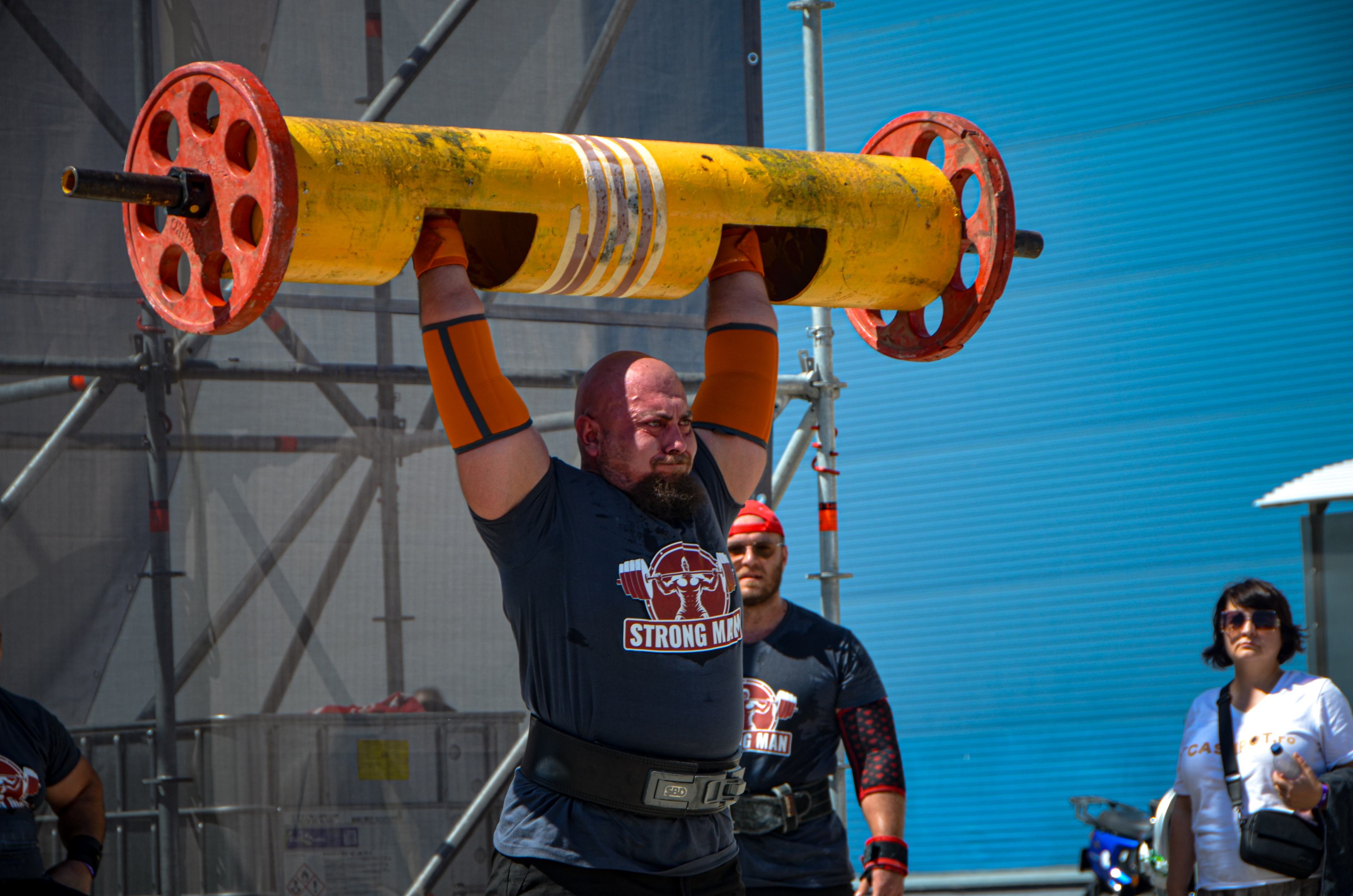
Muscle Under the Microscope - Episode 4: Actin and Myosin - The Microscopic Motor
In the first three episodes we moved from whole-muscle anatomy to the muscle fiber and then to the differences between fast and slow fibers in strongman. Now we reach one of the most important points in the entire series: the meeting of actin and myosin. Here, at a nanometer-scale level, chemical energy is converted into mechanical work. A person can see a barbell rise from the floor, but the mechanism that makes that movement possible is far smaller than the eye can resolve: myosin heads interact cyclically with actin filaments and use energy released from ATP to generate tension and filament sliding.
This is the microscopic motor of skeletal muscle contraction. It is not a simple one-piston engine, but a regulated molecular assembly in which calcium, troponin and tropomyosin control myosin access to actin, while the ATP-ADP-inorganic phosphate cycle changes the mechanical state of the myosin head. Modern structural and biochemical research confirms that actin-myosin interaction is the basis of force production in striated muscle.
For a strongman athlete, this biology is not a laboratory curiosity. Every kilogram moved, every second spent in a carry and every repetition depends on enormous numbers of molecular cycles operating in parallel. The better we understand this level, the easier it becomes to understand why strength, speed, fatigue and recovery are not separate phenomena.
Chapter 1 - What are actin and myosin?
Actin and myosin are contractile proteins, but they have different roles. Actin primarily forms the thin filaments of the sarcomere. Myosin forms the thick filaments and contains the motor domain capable of binding and hydrolyzing ATP. In skeletal muscle, myosin heads project toward actin filaments and can form cross-bridges when binding sites are available. Through repeated cycles of attachment, force production and detachment, actin is moved relative to myosin, shortening the sarcomere.
Actin should not be imagined as a passive rope. The actin filament is a dynamic structure organized together with tropomyosin and troponin, which participate in regulating myosin access. Myosin, in turn, is not merely structural material but a molecular motor with ATPase activity. Its motor domain uses ATP and its chemical transitions to change molecular conformation. This coupling between chemistry and mechanics is why myosin is considered a molecular motor.
Chapter 2 - The sarcomere - where the motor works
Actin and myosin do not float freely through the cell. They are organized into sarcomeres, the repeating contractile units of myofibrils. Thin actin filaments are anchored near Z discs and extend toward the center of the sarcomere. Thick myosin filaments occupy the central region. During contraction, the filaments slide past one another, increasing overlap without requiring the individual filaments themselves to shorten. This is the basis of sliding-filament theory.
It is important to separate sarcomere shortening from protein shortening. Actin and myosin are not like two rods that contract. Their length remains largely unchanged while their relative position changes. Myosin heads pull actin filaments toward the center of the sarcomere, and thousands of sarcomeres arranged in series contribute to the shortening of a myofibril, a fiber and ultimately the whole muscle.
Chapter 3 - The myosin head - the actual motor
The most fascinating part of myosin is its motor head. It contains a region that interacts with actin and a nucleotide-binding site for ATP and its hydrolysis products. Changes in the ATP state alter the conformation of the myosin head and prepare the molecule for another interaction. In muscle, the mechanical cycle is tightly coupled to the ATPase cycle.
A useful mental picture is a molecular hand that repeatedly grips, pulls and releases a track, but the analogy has limits. There is not one myosin head moving an entire filament. Within a sarcomere, a huge number of heads operate simultaneously or sequentially, and the distribution of their mechanical states determines the resulting tension. The force observed in a muscle is therefore the collective property of an enormous population of molecular motors.
Chapter 4 - ATP - the fuel that allows the cycle
ATP is the cell’s immediate energy currency and is essential to the actin-myosin cycle. A crucial point is that ATP is not used only to produce a force-producing movement. Binding of ATP to myosin promotes detachment from actin after a strongly bound state. ATP is then hydrolyzed, and the resulting energy is used to prepare the myosin head for the next stage of the cycle.
During a strongman event, total muscle ATP should not be viewed as a simple tank that the athlete empties. ATP is continuously consumed and resynthesized. Phosphocreatine, glycolysis and oxidative metabolism contribute in different proportions depending on intensity and duration. Therefore, the ability to produce force depends not only on the actomyosin motor, but also on the capacity of the entire system to fuel that motor.
Chapter 5 - The actin-myosin cycle step by step
We can follow the cycle in broad steps. First, ATP binds to myosin and promotes detachment from actin. Second, ATP is hydrolyzed to ADP and inorganic phosphate, and the myosin head enters a primed state. Third, if the binding site on actin is available, myosin can bind initially in a weakly bound state. Transitions then lead toward strongly bound states, and release of inorganic phosphate is associated with entry into a force-producing state. Later, ADP release followed by binding of a new ATP allows the cycle to continue. The detailed kinetics are more complex than this educational sequence, and modern models include several intermediate states.
The key idea is simple: each chemo-mechanical cycle allows a myosin head to move among states with different binding and force properties. Repeating these cycles across millions of heads produces measurable tension in the fiber. A maximal effort is, in this sense, a gigantic molecular orchestra.
Chapter 6 - Calcium - the switch that opens access to actin
If ATP powers the motor, calcium controls when the motor can act. In striated muscle, calcium binds to troponin C. This changes the conformation of the troponin-tropomyosin system on the thin filament and increases access to myosin-binding sites on actin. In this way, the electrical excitation signal is converted into a molecular condition favorable for contraction.
When calcium is removed from the cytosol and taken back up by the sarcoplasmic reticulum, tropomyosin returns toward a position that reduces myosin access to actin, and active force production declines. Relaxation is therefore not the absence of a process. It is an active process involving calcium reuptake and a return of the contractile apparatus toward a state with lower probability of actin-myosin interaction.
Chapter 7 - Why myosin does not pull actin like a continuous cable
A popular explanation says that a myosin head “pulls actin” and then repeats the same movement. That is useful as a starting point, but incomplete. The filaments experience a large number of stochastic interactions, and not all myosin heads are in the same state at the same moment. Some are detached, some weakly attached, and some strongly attached. Total tension arises from the distribution of these states and from the forces generated by the population. Modern literature describes a multi-state mechano-chemical cycle rather than one identical mechanical stroke repeated in isolation.
This perspective matters for understanding force. Force depends not only on the total number of myosin heads, but also on how many are in tension-producing states and how much force each can produce under the mechanical and biochemical conditions. Temperature, load, muscle length, pH, inorganic phosphate and calcium can alter contractile behavior. During fatigue, for example, changes in pH, phosphate and calcium can reduce force production at the myofilament level.
Chapter 8 - Actin, tropomyosin and troponin - the control system
Actin is the track on which myosin interacts, but the track has a controlled access system. Tropomyosin is associated with the actin filament and, at rest, contributes to blocking or reducing access to myosin-binding sites. Troponin is the regulatory complex that links the calcium signal to the change in this state. The components work together, not in isolation.
When calcium binds troponin C, conformational changes favor movement of tropomyosin and increase the probability that myosin will find available binding sites on actin. Once some heads bind strongly, actin-myosin interactions can also contribute to cooperative activation of the thin filament. The contractile apparatus therefore behaves as a coordinated system rather than millions of completely independent motors.
Chapter 9 - How the microscopic motor becomes force
To produce force at the muscle level, we have to pass through several organizational levels. One myosin head contributes one microscopic interaction. Many heads generate tension in a thick filament. Thin and thick filaments form a sarcomere. Sarcomeres form myofibrils, myofibrils fill muscle fibers, and fibers are organized into fascicles and the whole muscle. Force must then be transmitted through the cytoskeleton, extracellular matrix and tendon to bone. A limitation at any level can affect the final mechanical result.
That is why “how strong is myosin?” is not the same question as “how strong is the athlete?” Favorable myosin kinetics can contribute to high speed, but the athlete also needs sufficient contractile cross-sectional area, neural recruitment, favorable architecture and efficient force transmission. The microscopic motor is necessary, but it is not the whole vehicle.
Chapter 10 - Actin and myosin in maximal strongman strength
In a near-maximal deadlift, mechanical demand is so high that the system must generate very high tension. The nervous system recruits many motor units, and contractile activation rises within the recruited fibers. At the microscopic level, this means an enormous number of myosin heads enter cycles of interaction with actin. Each individual contribution is tiny, but their sum becomes sufficient to generate hundreds of kilograms of external force.
If the bar breaks slowly from the floor, it does not mean actin and myosin “cannot pull hard enough.” It means every link between the nervous system and the external load must produce and transmit enough mechanical moment. Muscle length, joint position, shortening velocity, moment arms and tendon properties influence how much of the microscopic force reaches the bar. Strongman turns microscopic biology into a gigantic mechanical problem.
Chapter 11 - Myosin speed and differences between fiber types
In the previous episodes we discussed type I, type IIa and type IIx fibers. One explanation for their different contractile speeds is the properties of their myosin isoforms. Different isoforms alter the kinetics of the actomyosin cycle and contribute to different shortening velocities. This is one reason MyHC type is such an important marker of contractile behavior.
Fast fibers do not have a single molecular button that says “produce maximum.” Their properties arise from the whole system: myosin isoform, the amount of myosin and actin, sarcomere organization, calcium handling, sarcoplasmic reticulum, mitochondria, metabolic enzymes and membrane properties. As a result, two fibers placed in the same broad classification can still differ meaningfully in function.
Chapter 12 - What happens to actin and myosin during fatigue?
Fatigue is not simply an abstract loss of energy. Changes occur at the myofilament level that can reduce force production. During intense exercise, changes in inorganic phosphate, hydrogen ions and calcium availability can affect myofibrillar calcium sensitivity and the probability of forming strongly force-producing states. The effect depends on conditions, temperature and fiber type.
For a strongman, this helps explain why the same weight can feel very different after several events. The muscle may still contain energy substrates and the athlete may still recruit motor units, but the contractile apparatus and its local chemical environment are no longer in the same state as during the first attempt. In competition, fatigue accumulates across several levels at once: neural, metabolic, contractile and mechanical.
Chapter 13 - When the motor gets stuck - rigor and the lack of ATP
An impressive detail of the actin-myosin cycle is that ATP is also required for myosin detachment from actin. When ATP is absent, strong binding can persist, producing the rigor state. The same fundamental principle is involved in rigor mortis, in which ATP resynthesis stops after death and actin-myosin cross-bridges can no longer be detached by the normal mechanism.
In a living athlete, of course, the situation is entirely different. ATP is continuously regenerated and the cycle continues as long as activation signals, calcium handling and energy supply allow it. The rigor example is useful precisely because it shows how fundamental ATP is to the microscopic dynamics of contraction.
Chapter 14 - Why this mechanism matters for strongman training
Training does not “train actin and myosin” as two isolated objects, but it changes the system in which they operate. Resistance training can increase fiber size and contractile protein content, alter myosin isoform expression and improve neural recruitment and coordination. At the same time, energy systems and oxygen-delivery systems adapt. The result is greater capacity to produce and sustain tension.
For strongman, this means good preparation must develop several links. Maximal strength exposes the system to very high tension. Explosive movements stress rapid force production. Carries and medleys develop the ability to repeat contractions under load and maintain technique under fatigue. At the microscopic level, all of these training forms converge in the same world: the interaction cycle between actin and myosin.
Chapter 15 - Conclusion - a motor only a few billionths of a meter across can move hundreds of kilograms
Actin is the thin filament on which myosin can generate force. Myosin is the molecular motor that uses ATP to cycle through attachment, force production and detachment. Calcium, troponin and tropomyosin regulate access to the interaction. The sarcomere organizes these elements, while millions of sarcomeres and millions of fibers transform microscopic events into visible contraction. This is the fundamental principle linking molecule to movement.
For strongman, the lesson is powerful: a record does not begin in the athlete’s hands or in the plates loaded onto the bar. It begins with a molecular interaction. The neural signal reaches the muscle fiber, calcium opens the path to actin, ATP fuels the myosin cycle, sarcomeres develop tension, the fiber transfers that tension into connective tissue, the tendon transmits it onward, and the whole system turns chemical energy into external movement.
When you watch a strongman lift a huge weight, what you see is only the final chapter. The real story begins much lower, inside the actin-myosin microscopic motor. And that motor works again and again, on an extraordinary scale, so that a single repetition can exist.
Scientific bibliography
1. Geeves MA, Holmes KC. The molecular mechanism of muscle contraction. Advances in Protein Chemistry. 2005;71:161-193.
2. Gordon AM, Homsher E, Regnier M. Regulation of contraction in striated muscle. Physiological Reviews. 2000;80(2):853-924.
3. Rayment I. The structural basis of the myosin ATPase cycle. Journal of Biological Chemistry. 1996;271(4):1583-1586.
4. Månsson A. Actomyosin interaction: from kinetics to mechanics. Cellular and Molecular Life Sciences. 2010;67:151-164.
5. Smith DA, Stehle R. Cross-bridge cycling in skeletal muscle: molecular and mechanical perspectives. Reviews and conceptual literature on muscle contraction and fatigue.
6. Myosin isoforms and the mechanochemical cross-bridge cycle. Journal of Experimental Biology. 2016. DOI: 10.1242/jeb.124594.
7. The cross-bridge cycle and skeletal muscle fatigue. Exercise and Sport Sciences Reviews. 2007;35(3):157-163.
8. Special Issue: The Actin-Myosin Interaction in Muscle - Background and Overview. International Journal of Molecular Sciences. 2019;20(22):5715. DOI: 10.3390/ijms20225715.
Editorial note: modern models of the actin-myosin cycle include more intermediate states than the simplified educational sequence used here. The relationships among calcium, troponin, tropomyosin, ATP, phosphate and the mechanical states of myosin are dynamic and depend on physiological and mechanical conditions.
More From The Journal

Training
The Science of Strongman Training - Episode X: Competition Programming
How to build a complete Strongman competition preparation block: calendar, events, volume, intensity, specificity, simulations, autoregulation, tapering and fatigue management.

Training
The Science of Strongman Training - Episode IX: Peaking
Peaking in Strongman: how to turn months of training into maximal competition performance by reducing fatigue, preserving adaptations and managing specificity.