
Physiology
Muscle Under the Microscope - Episode 1: The Microscopic Anatomy of Muscle
September 6, 2026
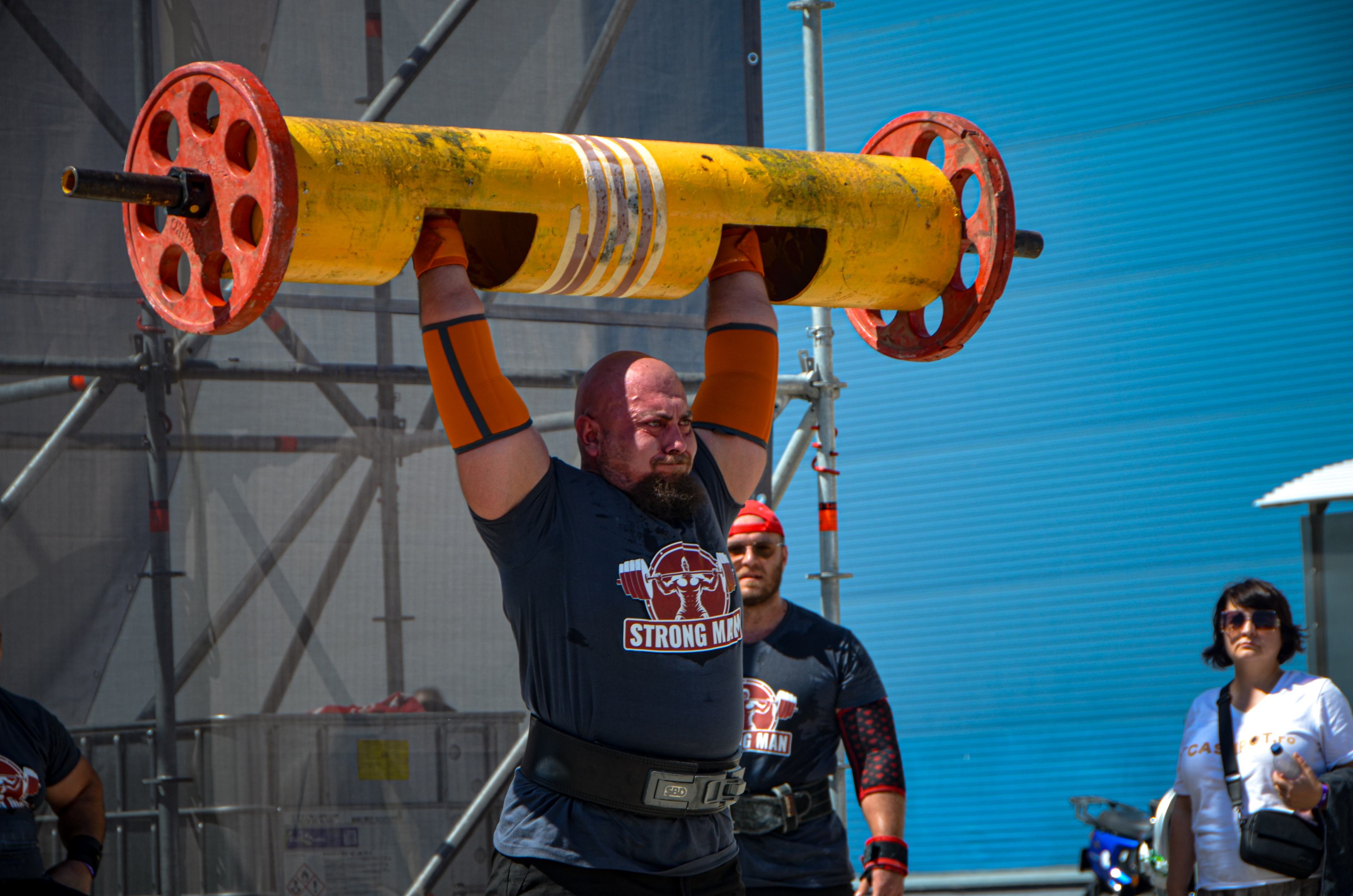
Muscle Under the Microscope - Episode 1: The Microscopic Anatomy of Muscle
When we look at a muscle from a distance, we see a compact organ. Under the microscope, that appearance disappears. Skeletal muscle is a hierarchical biological architecture in which connective tissue, muscle fibers, myofibrils, sarcomeres, membranes, sarcoplasmic reticulum, mitochondria and many structural proteins are integrated into a system capable of converting a neural signal into measurable mechanical force. This microscopic anatomy is the foundation of every muscular performance, from posture and walking to maximal strongman lifts.
The Muscle Under the Microscope series starts here. Before discussing hypertrophy, fatigue, adaptation or records, we need to understand the living material that makes those phenomena possible. In this first episode we move from the whole organ to the fascicle, from the fascicle to the fiber, from the fiber to the myofibril and from the myofibril to the sarcomere. Then we examine the molecular machinery of contraction and the infrastructure that keeps the contractile system mechanically integrated.
Chapter 1 - From organ to microscopic architecture
Skeletal muscle is a composite organ. The structures that generate force are mainly muscle fibers, but fibers do not operate in isolation. They are embedded in a connective-tissue system that forms the epimysium, perimysium and endomysium. These compartments organize fibers, provide mechanical support and create pathways for vessels and nerves. At the ends of the muscle, connective tissue continues into tendon and then into the structures that anchor the muscle. The muscle therefore should not be viewed as a contractile cable that suddenly ends at the tendon, but as a continuous force-transmission system.
The epimysium surrounds the muscle as a whole. Beneath it, bundles of muscle fibers are grouped and separated by perimysium. Each fiber is surrounded by endomysium, a fine network of extracellular matrix and connective tissue. In transverse sections this organization appears as compartments containing individual fibers. In longitudinal sections, fibers appear elongated and may run parallel to the tendon axis or at an angle, depending on muscle architecture. This geometry has mechanical consequences: pennation angle, fiber length and fascicle arrangement all affect how fiber shortening relates to whole-muscle force.
Chapter 2 - Connective tissue and the extracellular matrix
Muscle connective tissue is sometimes treated as simple packaging. Modern muscle biology shows that it is much more. The extracellular matrix contains collagens, glycoproteins, proteoglycans and other molecules that create a mechanical and biological environment for muscle fibers. The endomysium and perimysium stabilize fibers and fascicles and participate in tension distribution. The extracellular matrix is also a signaling platform: integrins and other membrane complexes link the outside of the cell to the cytoskeleton and contractile apparatus. Events inside the sarcomere can therefore be connected mechanically and chemically to the tissue outside the fiber.
Type I collagen is a major component of connective tissues that resist tensile loading. Other collagen types and associated molecules contribute to microarchitecture. The network is not static. Exercise, immobilization, aging, inflammation and injury can alter extracellular composition and organization. When fibrosis develops, excessive or reorganized extracellular material can change tissue mechanics and interfere with fiber function. Microscopic anatomy is therefore not a frozen photograph, but a living landscape that continually adapts.
Chapter 3 - The muscle fiber - the giant force-producing cell
A skeletal muscle fiber is a highly specialized cell formed during development through the fusion of myogenic precursor cells. The result is a very long multinucleated cell. Nuclei are characteristically positioned at the periphery, immediately beneath the sarcolemma. Dimensions vary with muscle and individual, but human fiber diameters are commonly in the tens of micrometers and lengths can reach millimeters or more. This geometry explains why the cell needs sophisticated internal infrastructure to move signals and resources rapidly.
The sarcolemma is the muscle-fiber membrane. It separates sarcoplasm from the extracellular environment and contains proteins involved in excitability, adhesion and force transmission. The sarcoplasm contains very large numbers of myofibrils, mitochondria, glycogen particles, lipid droplets, sarcoplasmic reticulum and other structures. There is no single metabolic center feeding the entire fiber uniformly. The local distribution of mitochondria, reticulum and membrane networks brings energy and ion-handling capacity close to regions of demand.
Chapter 4 - Myofibrils - the contractile framework of the fiber
Myofibrils are cylindrical structures aligned along the length of the fiber. They occupy a large fraction of cellular volume and are built from repeated sarcomeres. The striated appearance of skeletal muscle seen in light microscopy is a direct expression of this regular organization. In mature fibers, sarcomeres in neighboring myofibrils are aligned transversely, so Z lines appear in register. This geometry allows many contractile units to contribute coherently to force.
Functionally, a myofibril is a series of tension-producing units, but it is important not to isolate it conceptually from the rest of the cell. Myofibrils are connected to one another and to the membrane through cytoskeletal proteins. When a sarcomere produces tension, that tension must be distributed to neighboring myofibrils and then to the sarcolemma and extracellular matrix. A key feature of muscle is therefore summation: extremely small molecular forces become large tissue-level forces when huge numbers of structural elements act in parallel and are mechanically connected.
Chapter 5 - The sarcomere - the microscopic unit of contraction
The sarcomere is the fundamental structural and functional unit of the myofibril. It extends from one Z line to the next. Inside are thin actin filaments and thick myosin filaments arranged with high precision. Electron microscopy reveals the A band, I band, H zone and M line. The A band corresponds mainly to the length of the thick filaments. The I band is the region dominated by thin filaments, the H zone is the central region with little or no thin-filament overlap in the relaxed sarcomere, and the M line lies at the center.
Contraction is explained by the sliding-filament mechanism. Actin and myosin do not shorten like solid rods. Instead, myosin heads repeatedly interact with actin and pull the thin filaments toward the center of the sarcomere. Z lines move closer together, the sarcomere shortens and the muscle develops tension. The relationship between sarcomere length and filament overlap affects force potential. Too little overlap or excessive crowding can reduce the number of productive actin-myosin interactions.
Chapter 6 - Actin, myosin, troponin and tropomyosin
Myosin is the molecular motor of contraction. Thick filaments contain many myosin molecules, each with a globular head capable of binding actin and using ATP energy. Actin forms thin filaments, while tropomyosin and the troponin complex regulate their interaction with myosin. In resting muscle, the position of tropomyosin restricts effective actin-myosin engagement. Calcium changes that state.
When cytosolic Ca2+ rises, calcium binds troponin C. The troponin complex changes conformation, tropomyosin moves and actin binding sites become available. Myosin then enters a cycle involving ATP binding, ATP hydrolysis, actin attachment, product release and detachment after binding a new ATP molecule. Repetition of this cycle on a massive scale produces filament sliding and muscle tension. The energy is not created by actin; it is supplied by cellular metabolism to sustain the molecular cycle.
Chapter 7 - Titin, desmin and the contractile cytoskeleton
A muscle could not function if actin and myosin were its only mechanical components. Titin is one of the largest known proteins and extends within the sarcomere from the Z line toward the thick filament. It contributes to filament positioning, passive elastic behavior and sarcomere stability. When muscle is stretched, titin elasticity contributes to resistance against extension and to the tendency to return toward shorter lengths.
Desmin is an intermediate filament that helps align myofibrils and connects Z-line regions with the cytoskeleton and membrane. Other proteins organize links between the contractile apparatus and the sarcolemma. Together, these systems form a mechanical distribution network. Contraction is therefore not merely the internal sliding of two filament systems. Force must be directed, distributed and absorbed without mechanically destroying the cell.
Chapter 8 - The sarcolemma, costameres and the connection to the outside
The sarcolemma is more than an electrical boundary. It belongs to a mechanical system connecting the internal cytoskeleton to the extracellular matrix. Costameres are protein assemblies found especially near Z lines and connect actin, cytoskeletal proteins, membrane proteins and extracellular components. Through this network, some force can be transmitted laterally rather than only along the long axis of the fiber.
Dystrophin is a classic example. It is part of a complex linking the cytoskeleton to the membrane and extracellular matrix. When dystrophin is absent or severely defective, the sarcolemma becomes vulnerable to repeated mechanical stress and progressive fiber damage can occur. Duchenne muscular dystrophy demonstrates clinically how important this microscopic linkage is. A protein that does not generate contraction directly can still be essential for allowing muscle to survive contraction.
Chapter 9 - T tubules and the sarcoplasmic reticulum
A muscle fiber can have a diameter of tens of micrometers and a substantial length. If electrical activation stayed only at the surface, deep regions of the cell could not be activated quickly and uniformly. Transverse tubules, or T tubules, solve this problem. They are invaginations of the sarcolemma that penetrate between myofibrils and carry changes in membrane potential deep into the fiber.
In skeletal muscle, one T tubule is associated with two terminal cisternae of the sarcoplasmic reticulum, forming a triad. The sarcoplasmic reticulum is specialized for calcium storage and release. Depolarization of the T-tubule membrane activates CaV1.1, also known as the dihydropyridine receptor. CaV1.1 communicates functionally with ryanodine receptor 1, RyR1, in the sarcoplasmic reticulum. RyR1 releases Ca2+ into the sarcoplasm, directly linking the electrical event to the contractile machinery.
After activation, calcium must be removed rapidly to permit relaxation. SERCA pumps use ATP to return Ca2+ to the sarcoplasmic reticulum. Relaxation is therefore also an active energy-consuming process. At microscopic level, rapid contraction is the result of coordination among membranes, channels, pumps, calcium and myofilaments.
Chapter 10 - Mitochondria, glycogen and ATP economy
ATP is the immediate energy currency of muscle, but the amount stored at any moment is small relative to the demand of hard exercise. ATP therefore has to be resynthesized continuously. Phosphocreatine provides rapid ATP buffering, glycolysis can produce ATP from carbohydrates, and mitochondrial oxidative phosphorylation supports sustained production.
Under the microscope, mitochondria are not randomly distributed. They occur between myofibrils and close to the membrane and other regions of high energy demand. Glycogen is stored as granules in the sarcoplasm and can be mobilized for energy production. Different fiber types have different mitochondrial content, oxidative enzyme profiles, capillary supply and substrate-use patterns, but no human fiber is simply "aerobic" or "anaerobic." During maximal effort, energy systems overlap and contribute together, with their relative contributions changing over time.
Chapter 11 - Capillaries and muscle microcirculation
A dense capillary network lies around muscle fibers. These vessels allow exchange of oxygen, nutrients and metabolic products. The small distance between blood and sarcoplasm supports rapid diffusion. Fibers with higher oxidative capacity are generally associated with greater capillary density, helping sustain aerobic metabolism.
During intense exercise, the situation becomes highly dynamic. Muscle contraction raises tissue pressure and can temporarily compress vessels, especially when intramuscular tension is very high. Between contractions, perfusion can increase and deliver oxygen and substrates. This interaction between compression and reperfusion helps explain why a muscle working at very high intensity can rapidly become dependent on its local energy stores.
Chapter 12 - Muscle fiber types
Human skeletal muscle is commonly described in terms of fibers expressing myosin heavy-chain isoforms associated with types I, IIA and IIX. Type I fibers generally contract more slowly and have greater oxidative capacity and fatigue resistance. Type II fibers are faster and can support high power output. These labels should not be treated as exact synonyms for "endurance" and "explosiveness." Real fiber behavior depends on many components, including fiber size, mitochondrial content, metabolic enzymes, capillarization and muscle architecture.
Human muscle also contains hybrid fibers that coexpress more than one myosin heavy-chain isoform. Fiber phenotype is plastic. Training and habitual use can shift some contractile and metabolic characteristics. In strongman, this matters because events demand combinations of qualities: a maximal lift can require high force and rapid neural activation, while carries or repeated efforts require the ability to sustain force under accumulating fatigue.
Chapter 13 - Myonuclei and satellite cells
The presence of multiple nuclei in a muscle fiber is a functional necessity. A very long, large cell has to regulate RNA and protein synthesis across a broad cytoplasmic volume. Peripheral myonuclei contribute to gene regulation and maintenance of local fiber domains. Their spatial relationship to the contractile apparatus is part of functional muscle organization.
Satellite cells lie between the sarcolemma and basal lamina. These muscle stem cells can be activated after injury and, in some contexts, by mechanical loading. They can proliferate, differentiate and contribute nuclei to existing fibers, supporting repair and remodeling. Hypertrophy should not be reduced to the claim that every increase in muscle size is caused exclusively by satellite cells. Adult fibers can enlarge through accumulation of contractile proteins and structural remodeling, while the importance of new myonuclei depends on the stimulus and biological context.
Chapter 14 - How microscopic anatomy is studied
Light microscopy is used to evaluate general architecture. Muscle sections can reveal fibers, nuclei, fascicles and features of connective tissue. Histological stains help distinguish tissue components, while enzyme histochemistry and immunohistochemistry can provide information about metabolism and specific proteins. In muscle pathology, frozen sections and panels of histochemical techniques have substantial diagnostic value.
Electron microscopy provides much higher resolution. Sarcomeres, Z lines, sarcoplasmic reticulum, T tubules and mitochondria can be examined at ultrastructural scale. Immunofluorescence and confocal microscopy can localize proteins in relation to cellular structures, while modern imaging approaches can investigate molecular assemblies with extraordinary precision. Each method reveals a different layer of the same biological puzzle.
Chapter 15 - Why microscopic anatomy matters in strongman
In strongman, we see the outcome: a load is lifted, carried, pressed, pulled or stabilized. At microscopic level, however, we see the machinery. A heavy object moves because millions of sarcomeres generate tension, fibers are organized into a useful geometry, calcium is released and recovered rapidly, ATP is continuously resynthesized, and connective tissue transmits force toward tendon and bone. Performance is therefore an emergent property of a biological architecture that begins at the molecular level.
We should also avoid the opposite oversimplification. A larger muscle cross-sectional area can increase force potential, but it does not determine performance by itself. Voluntary force also depends on motor-unit recruitment, discharge rate, coordination, internal muscle architecture, sarcomere length, tendon properties and technique. Microscopic anatomy is the foundation of the building, while neural control, biomechanics and physiology determine how the building behaves.
Bibliography
1. Shishmarev D. Excitation-contraction coupling in skeletal muscle: recent progress and unanswered questions. Biophysical Reviews. 2020;12:143-153. DOI: 10.1007/s12551-020-00610-x.
2. Berchtold MW, Brinkmeier H, Muntener M. Calcium ion in skeletal muscle: its crucial role for muscle function, plasticity, and disease. Physiological Reviews. 2000;80(3):1215-1265.
3. Lieber RL, Ward SR. Skeletal muscle design to meet functional demands. Philosophical Transactions of the Royal Society B. 2011;366:1466-1476.
4. Gillies AR, Lieber RL. Structure and function of the skeletal muscle extracellular matrix. Muscle & Nerve. 2011;44(3):318-331.
5. Frontera WR, Ochala J. Skeletal muscle: a brief review of structure and function. Calcified Tissue International. 2015;96:183-195.
6. Schiaffino S, Reggiani C. Fiber types in mammalian skeletal muscles. Physiological Reviews. 2011;91(4):1447-1531.
7. Murach KA, Bagley JR. Skeletal muscle hypertrophy with resistance training: a review of research mechanisms and implications. Sports Medicine. 2022;52:1-15.
8. Keefe AC, Kardon G. A new role for the satellite cell: skeletal muscle regeneration and beyond. Development. 2014;141:1943-1951.
9. Squire JM. Architecture and function in the muscle sarcomere. Current Opinion in Structural Biology. 2016;37:56-61.
10. Al-Qusairi L, Laporte J. T-tubule biogenesis and triad formation in skeletal muscle and implication in human diseases. Skeletal Muscle. 2011;1:26.
11. Dubrulle J, et al. Structure-function relationships in the skeletal muscle extracellular matrix. Journal of Physiology. 2023.
12. Murach KA, Bagley JR. Fiber-type traps: revisiting common misconceptions about skeletal muscle fiber types. Journal of Physiology. 2024.
Editorial note: the bibliography combines reviews and reference works used to verify the scientific content. Terminology has been adapted for a broad audience while preserving physiological precision.
More From The Journal

Training
The Science of Strongman Training - Episode X: Competition Programming
How to build a complete Strongman competition preparation block: calendar, events, volume, intensity, specificity, simulations, autoregulation, tapering and fatigue management.

Training
The Science of Strongman Training - Episode IX: Peaking
Peaking in Strongman: how to turn months of training into maximal competition performance by reducing fatigue, preserving adaptations and managing specificity.