
Physiology
Bones, Tendons and Ligaments: Structures That Must Withstand Force - Episode 6: Ligaments and Joint Stability
September 6, 2026
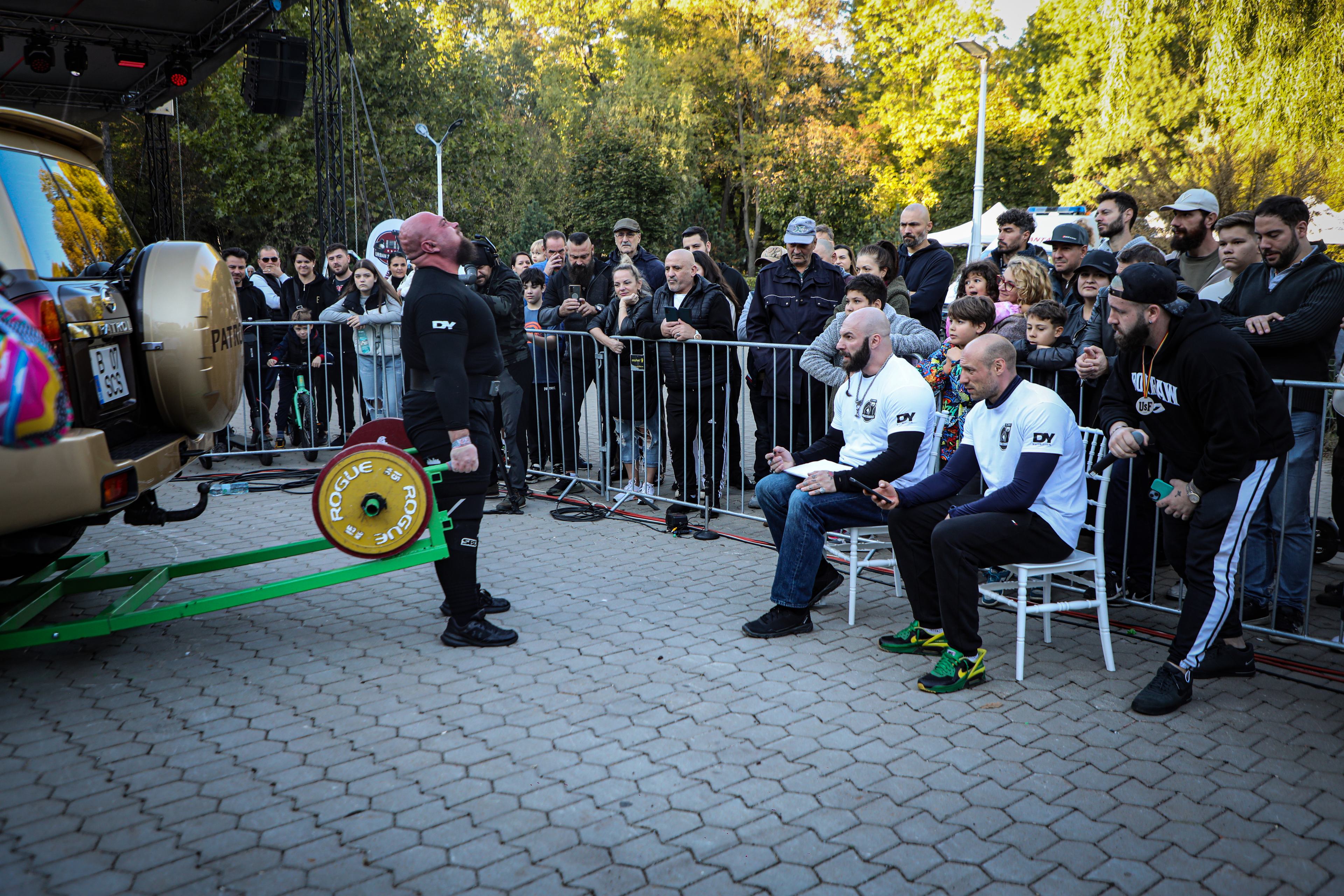
1. Introduction: ligaments, stability, and the problem of extreme force
In Strongman, a joint is never simply the place where two bones meet. It is a mechanical node where muscle forces, ground reactions, implement mass, acceleration, braking, and control strategies converge. Ligaments are part of the architecture that allows those forces to be managed without uncontrolled movement. They limit excessive excursions, guide relationships between joint surfaces, and contribute sensory information that helps the nervous system maintain position. In a sport where implements can weigh hundreds of kilograms and where one event may combine lifting, walking, rotation, shifting center of mass, and fatigue, stability becomes a performance requirement rather than only a medical concern.
The ligament is often imagined as a passive band holding bones together. The image is useful but incomplete. Ligament biology is active: cells respond to mechanical stimuli, extracellular matrix is remodeled, and tissue properties change over time. Ligaments also work with other structures. A knee collateral ligament shares load with muscle and capsule; an ankle ligament is influenced by calf and foot control; shoulder stability depends on capsule, labrum, rotator cuff, and scapular position. True stability is distributed across a system.
For a strength athlete, an important question is how much stability is enough. The answer cannot simply be as much as possible. A completely rigid joint would lose useful mobility, while excessive laxity would require more neuromuscular work and could permit unwanted motion. Optimal biomechanics is a dynamic compromise between freedom and precise restraint. Ligaments are one of the structures that create that compromise at very small scales.
1.1. Why a Strongman athlete needs stability before maximal force
External force must pass through joints before it reaches the ground or the implement. When a joint loses control, some force is spent on corrections instead of propulsion. In competition, the difference between a stable trajectory and one that requires constant correction can become seconds, repetitions, or distance. Repeated changes in trajectory can also change which tissues receive stress. Stability is therefore a problem of mechanical efficiency as well as protection.
1.2. A stable joint is a joint that can move
Stability should not be confused with absence of movement. A healthy knee, shoulder, or ankle moves normally. The goal is controlled excursion rather than blocked motion. During a carry, for example, the ankle has to adapt to each ground contact, the knee must allow progression, and the hip must move the center of mass forward. Ligaments help keep these movements within functional mechanical windows.
2. The ligament as living tissue: anatomy and organization
Ligaments are connective tissue structures specialized for transmitting and controlling tensile forces. Their dominant component is extracellular matrix rich in collagen, especially type I collagen. Collagen fibers are arranged into fascicles, and the fascicles form an architecture that reflects loading directions. Water, proteoglycans, and other matrix components are present between the structural elements. Ligament cells, especially fibroblasts and specialized forms, produce and remodel this matrix.
2.1. Collagen and hierarchical architecture
Type I collagen provides high tensile strength. Molecules assemble into fibrils, fibers, and fascicles, allowing stress to be distributed. Fibers can have microscopic waviness, often called crimp, which decreases early in loading. As load rises, more fibers are progressively recruited. This explains why the ligament can be relatively compliant early in motion and become much stiffer as loading increases. Not every structural element becomes taut at the same moment.
2.2. Fascicles and load sharing
Within a complex ligament, fascicles can be recruited differently depending on joint angle. One fascicle may be highly loaded in one position and much less loaded in another. This makes stability position-dependent. For the athlete, the same external weight does not necessarily create the same ligament demand at different joint angles. Muscles can also change joint relationships and therefore change how much load reaches passive structures.
2.3. The enthesis and bone attachment
Ligaments attach to bone through specialized regions called entheses. The properties of soft tissue and mineralized bone change gradually across this interface. The transition reduces abrupt stress concentration and permits force transfer. During injury, location matters: damage in the ligament body can behave differently from damage near the insertion. Clinical assessment benefits from knowing exactly where the structure is affected.
3. Ligaments and static versus dynamic stability
Passive stability is provided by bone geometry, capsule, ligaments, menisci, and labrum depending on the joint. Active stability is generated mainly by muscles and tendons and regulated by the nervous system. During a heavy effort these components work at the same time. Ligaments do not simply take over when muscle works less, and muscle cannot erase the mechanical role of a ligament. The joint is stabilized by shared contribution.
3.1. Primary and secondary stabilizers
Some structures are called primary stabilizers because they provide the major resistance against a particular motion. Others act as secondary stabilizers and become more important in specific positions or when the primary structure is insufficient. The distinction is useful but not absolute. In the knee, thigh muscles, menisci, capsule, and ligaments share the task according to load and angle. In the shoulder, capsule and ligaments work with the rotator cuff and scapular control.
3.2. Stability during loaded walking
Farmer's walk and yoke walk are excellent examples of dynamic stability. With every step the center of mass moves, and the joints must absorb and redirect force. If the implement swings, additional corrections are required. Ligaments help limit the amplitude of these excursions while muscles generate the active correction. Efficient carrying is therefore partly an exercise in repeated stability control.
4. The force-deformation curve: how a ligament responds to load
When a ligament is loaded, the relationship between force and deformation is not perfectly linear from the beginning. Wavy collagen fibers progressively align, after which the tissue enters a stiffer region. At high deformation, microscopic damage begins, and continued loading may produce structural failure. This is important for understanding injury because tissue damage does not begin only at the moment a complete rupture becomes visible. Microstructural damage may occur first.
4.1. Stress, strain, and material properties
Stress can be expressed as force relative to cross-sectional area, while strain describes relative change in length. These concepts separate material behavior from total structure size. A thicker ligament can tolerate more absolute force without necessarily having a stronger material. Conversely, a smaller structure may contain highly organized tissue but still have lower absolute capacity. This distinction is essential in biomechanical research.
4.2. Stiffness and compliance
Stiffness describes resistance to deformation, while compliance describes how readily a structure deforms. Neither is universally good or bad. Some compliance permits movement and energy absorption, while sufficient stiffness supports control. A high-level strength athlete needs a system that can become appropriately stiff during force production while retaining enough mobility to execute efficient technique.
4.3. Viscoelasticity and time dependence
Ligaments are viscoelastic. Under constant load, deformation may gradually increase, a phenomenon called creep. At constant length, internal stress may decrease over time, called stress relaxation. These behaviors explain why tissue responds differently to a long hold than to a brief load. In training, time under tension and repeated positions can change the acute response without automatically implying permanent damage.
5. Anisotropy: why force direction changes the problem
Ligaments are anisotropic, meaning their properties depend on loading direction. Collagen fibers are oriented along preferred mechanical axes established through development and adaptation. When force aligns with this architecture, resistance is efficient. When a movement combines rotation, shear, and translation in an unexpected way, tissue loading becomes more complex. This matters for unusual implements and unpredictable movements.
5.1. Valgus and varus
Valgus and varus describe frontal-plane angulation. In the knee, the collateral ligaments help control these motions. Real movement, however, is rarely isolated to one plane. A knee moving into valgus while also rotating experiences a different loading pattern from slow, pure valgus. This is one reason injury mechanism is such a valuable part of clinical assessment.
5.2. Rotation and translation
Rotation and translation can occur together. In controlled lifting they are usually small and coordinated. During a sudden perturbation, excursion can increase very quickly. Ligaments help limit these changes. If external force exceeds the combined ability of tissue and muscular control to manage the movement, injury can occur.
6. Proprioception: ligaments are also part of the sensory system
Ligaments contain sensory nerve endings and mechanoreceptors that contribute to information about joint state. These signals are integrated with input from muscle, tendon, skin, vestibular organs, and vision. There is not one single ligament sensor that dictates joint position. There is a distributed sensory system, and ligaments are part of it. After injury, mechanical and sensory changes can alter how the athlete perceives and controls the joint.
6.1. Mechanoreceptors and motor responses
Mechanoreceptors respond to characteristics of deformation and movement. Their information contributes to neural processing and can influence muscle activation. In fast tasks, this input is integrated with signals from other tissues. It is therefore inaccurate to say that a ligament sends a complete reflex by itself, but it is accurate to say that ligament sensory input can participate in neuromuscular control.
6.2. Why an injured joint can feel unstable
Perceived instability can arise from a combination of mechanical laxity, pain, altered sensory feedback, and protective motor strategies. An athlete may still be very strong while instinctively avoiding certain positions. Rehabilitation therefore has to restore more than strength. It must also restore control, tolerance, and confidence in movement.
7. The knee: a multi-planar stability system
The knee is one of the joints in which ligament function has been studied extensively. Cruciate and collateral ligaments work with menisci, capsule, and thigh muscles to control translation and rotation. In Strongman, these demands appear in squats, deadlifts, carries, sled work, loading, and movement under load. Even apparently vertical movement contains frontal and transverse components.
7.1. The anterior cruciate ligament
The anterior cruciate ligament contributes to limiting anterior tibial translation and controlling rotation. Injury can result from combinations of deceleration, rotation, and multi-planar loading. Strongman does not always reproduce field-sport mechanisms, but an athlete can still encounter risk when a loaded foot is planted, the body rotates, and an implement perturbs balance. ACL management should be individualized and rehabilitation is central.
7.2. The posterior cruciate ligament
The posterior cruciate ligament primarily limits posterior tibial translation. Evaluation of an injured PCL has to consider the entire knee system. Being able to squat is not sufficient evidence that the joint is ready for sport. Walking, unilateral control, range of motion, strength, and response to sport-like loading all matter.
7.3. The collateral ligaments
The medial collateral ligament and lateral complex contribute to resistance against valgus and varus as well as rotational control. During carries, the implement can create lateral oscillations that alter loading. Hip and foot function influence these motions, so a knee cannot be interpreted in isolation. Stability is a kinetic-chain phenomenon.
35. Joint stability as an emergent property of the whole system
When stability is discussed, it is tempting to choose one responsible structure: the ligament, the muscle, or the capsule. In reality, stability is an emergent property. It arises from bone geometry, tissue elasticity, ligament tension, muscular force, and the way the nervous system combines available information. During a demanding event, these components change simultaneously. Two anatomically similar joints can therefore respond differently to the same load when their training history, injury history, or neuromuscular adaptation differs.
35.1. Bone geometry versus ligament restraint
Some joints have more inherent bony stability. The hip is an example because the femoral head is deeply contained by the acetabulum. The shoulder sacrifices some bony congruence for mobility. Within one joint, geometry can also determine when a ligament becomes taut. Stability is therefore a property of the relationship between tissue and geometry, not of tissue in isolation.
35.2. Stability and freedom of movement
An athlete needs enough mobility to reach mechanically useful positions. If mobility is limited, compensation can occur elsewhere. If mobility is large and poorly controlled, passive excursion may increase. Effective training does not maximize one quality in isolation. It develops a useful relationship among range of motion, control, and load.
36. Ligaments and force transfer between body segments
Muscle force must reach the implement and the ground through a chain of segments. Ligaments help maintain the relative relationships among those segments. If the knee moves excessively during a squat, part of the force is redirected. If the shoulder does not maintain a controlled humeral center, pressing becomes less efficient. If the ankle collapses, ground reaction is transmitted differently. Stability is therefore a condition for mechanical continuity.
36.1. The kinetic chain in floor lifts
In the deadlift, force is transmitted from the foot through ankle, knee, and hip to the pelvis and trunk, then through the arms to the bar. Each region's ligaments contribute to relative segment control. If one segment deviates, others must compensate. A strong deadlift is therefore also a lift in which the joints remain stable enough to transfer energy without unnecessary loss.
36.2. The kinetic chain in carries
Carries make the kinetic chain cyclic. Every step creates another opportunity for a small deviation. If the foot lands poorly, knee and hip must correct. If the implement swings, shoulder and trunk respond. The longer the course, the more corrections accumulate. Ligament stability is therefore involved not in one moment, but across a long sequence of small mechanical decisions.
37. Stability and movement speed
Slow movement allows more time to observe and correct position. Fast movement reduces the time available for control. Event speed can be a competitive advantage, but it also requires greater neuromuscular capacity. Ligaments may respond differently to rapid loading because viscoelastic tissues have less time to redistribute stress, while muscles have to anticipate the movement. Velocity progression should therefore be treated as a separate progression rather than an accidental by-product of adding weight.
37.1. Acceleration at the start of an event
A rapid start of a farmer's or yoke changes system inertia quickly. The feet have to produce force while the kinetic chain permits acceleration. An athlete who is not prepared for that speed may take steps that are too large or lose alignment. Ligaments can then be exposed to movement patterns that were not present during slow lifting with the same weight.
37.2. Deceleration and stopping
Stopping a heavy object is not a trivial task. Body and implement have momentum, and the system has to dissipate it. Knee, ankle, hip, and trunk absorb braking forces. If the athlete stops with a sudden uncontrolled movement, loading may rise rapidly. In competition, deceleration can be as demanding as acceleration.
38. Stability under central and peripheral fatigue
Fatigue can have central and peripheral components. Regardless of mechanism, the effect on technique can include reduced force and reduced coordination. A very fatigued athlete may continue moving a load but use a different strategy. Ligaments are affected indirectly because muscles may become less effective at active joint control. This explains why a load tolerated at the start of a session can produce much less stable mechanics at the end.
38.1. Local fatigue
Local fatigue appears when certain muscles can no longer generate the same force or precision. If grip is the weak link, the implement may rotate and the athlete may change shoulder position. If the quadriceps fatigue, knee trajectory can change. If trunk extensors fatigue, posture may shift. Each compensation changes load distribution.
38.2. General fatigue and programming decisions
A training plan should not treat every day as though the athlete starts in identical condition. After competitions, travel, poor sleep, and hard blocks, control may be reduced. In those situations, reducing an event is not necessarily lost progress. It can preserve mechanical quality and reduce the chance that fatigue becomes an unnecessary exposure.
39. Ligament injury and factors that cannot be fully controlled
No strategy can eliminate all injuries. Individual anatomy, prior injury, surface, equipment, object contact, and unpredictable events are part of sport. The realistic goal is to reduce modifiable risk and increase capacity. Saying that an athlete should never get injured if they warm up properly is a simplification. Warm-up, strength, and technique may help, but they cannot provide absolute protection.
39.1. Previous injury history
A previous injury can change mechanics and confidence in a joint. Recurrence risk depends on many variables and differs by region. For an athlete with recurrent ankle sprains, unilateral control and graded exposure to direction changes may be important. For someone with shoulder instability, overhead positions should be restored through targeted progressive work.
39.2. Surface and environment
Competition surfaces change ground reaction and predictability of support. Strongman events can occur on rubber, concrete, grass, wood, or other materials. Outdoor conditions add temperature and moisture variability. Adapting to the environment is part of event preparation, and gradual exposure to unfamiliar surfaces can be useful.
40. Diagnosing a ligament problem
Diagnosis begins with history and mechanism, continues with clinical examination, and may include imaging. A clinician may assess laxity, tenderness, range of motion, and function. Radiography can be important when bone injury must be excluded, while MRI can help evaluate many soft tissues. Not every ligament complaint requires advanced imaging, but persistent symptoms should not be dismissed simply because the athlete is strong.
40.1. Clinical examination
Clinical tests are designed to provoke or demonstrate particular motions and assess laxity or pain. Their results have to be interpreted in context because sensitivity and specificity vary. The clinician often compares the affected side with the other side and combines several tests. One positive test is not equivalent to a complete diagnosis.
40.2. Imaging
Imaging can show ligament continuity, associated injury, edema, or bone changes. MRI is particularly useful for many soft tissues, but images must be correlated with symptoms and examination. A structural finding is not automatically the source of pain. Athletes can have imaging abnormalities that are not clinically important.
41. Neuromuscular rehabilitation and joint control
After ligament injury, rehabilitation must restore the function of the whole system. Strength work rebuilds muscular capacity, but control may also require balance tasks, directional changes, perturbations, and unilateral loading. For Strongman, these elements should eventually be connected to real implements. An athlete who can balance on one leg without load may still need significant progression before a heavy farmer's carry.
41.1. Proprioception and balance
Balance exercises challenge integration of sensory signals and muscular responses. They can be progressed by reducing the base of support, moving the head, adding perturbations, or introducing external loads. The purpose is not to turn rehabilitation into unstable-surface entertainment. The purpose is to train control that is relevant to the next demand.
41.2. Unilateral strength
Unilateral exercises can reveal strength and control differences hidden during bilateral movements. Split squats, step-ups, and selected carry variations can expose one limb at a time. They do not perfectly reproduce competition but can provide a bridge between rehabilitation and sport demand.
42. Periodization and passive tissues
Periodization can also be understood as tissue management. A base phase may allow more moderate volume and highly controllable movements. As competition approaches, event specificity and intensity rise, while total volume may fall to allow recovery. This protects not only the nervous system and muscle but also connective tissues that adapt more slowly.
42.1. Overlap among stimuli
Problems arise when several exercises create the same tissue demand. Squats, yoke, and sled work differ, but all load the lower limb. Log, axle, rows, and grip work can overlap at the elbow and shoulder. Periodization must consider these overlaps rather than only muscle-group splits.
42.2. The taper before competition
Tapering usually reduces volume while maintaining useful intensity so fatigue falls. For ligaments, the advantage is that the athlete reaches competition with fewer recent loading cycles and better neuromuscular control. The final week is not the time to magically strengthen a ligament. It is the time to avoid creating unnecessary fatigue or irritation immediately before competition.
43. What realistic injury prevention means in Strongman
Prevention is not a promise of an injury-free career. It is reduction of modifiable risk and improvement of capacity. It includes progression, technique, strength, control, recovery, appropriate equipment, and adaptation to event conditions. It also includes recognizing problems early and changing training before small problems become major injuries. That is training intelligence, not avoidance of hard work.
43.1. Primary prevention
Primary prevention begins before injury appears: building strength, control, and event tolerance. Movements and velocities are introduced gradually, and the athlete is not thrown directly into maximal competition stress. The goal is for mechanical novelty to be managed in training rather than discovered during the first event attempt.
43.2. Secondary prevention after injury
After injury, recurrence prevention includes understanding contributing factors, completing rehabilitation, and progressively exposing the athlete to the mechanism that previously caused the problem. A recurrent ankle sprain needs more than rest. A shoulder with instability needs control in positions relevant to pressing. The goal is to reduce the gap between current capacity and sport demand.
44. Biology, biomechanics, and the psychology of return
Return from ligament injury is not purely biological. An athlete may have a healing tissue and still avoid the position that caused the injury. Fear can alter muscle recruitment and technique. Excess confidence can push progression too quickly. Good recovery builds evidence, step by step, that the athlete can again control the required load.
44.1. Trust in the joint
Trust is not the same as courage. It can be built through repeated successful exposure. If an athlete progresses from 50% to 70% and then to 80% of a demand while maintaining control, the brain receives repeated evidence that the movement is possible again. Progression therefore has a psychological dimension as well as a mechanical one.
44.2. Fear and excessive protection
Overprotective movement can create excessive stiffness and loss of useful range. An athlete may brace the trunk too aggressively, shorten movement, and create compensations elsewhere. Rehabilitation should progressively restore controlled freedom. The aim is safety through capacity rather than safety through permanent avoidance.
45. Synthesis: where anatomy and Strongman practice meet
Anatomy tells us what structures exist. Biomechanics tells us how they are loaded. Physiology tells us how they respond. Programming tells us how often the exposure is repeated. In Strongman, all four need to connect. A healthy ligament is valuable, but it cannot compensate for poor technique and chronic fatigue. Very strong muscles are useful, but they cannot eliminate the role of ligaments. Efficient technique reduces mechanical waste, but it does not make the sport risk-free. The final result is produced by the whole system.
That leads to the central idea of this chapter: joint stability is the invisible infrastructure of strength. When it works well, the athlete does not think about it. When it is compromised, every step, clean, and press becomes a control problem. For an athlete seeking a long career, the goal is not to eliminate every load. It is to build capacity that can absorb varied demands and recover between them.
46. Extended conclusion: ligaments and the art of staying intact under enormous force
Ligaments are some of the quietest structures in the musculoskeletal system, but their quietness should not be mistaken for insignificance. They limit translation and rotation, guide motion, distribute load, and contribute to proprioceptive information. In a sport where objects are heavy, shapes change, and fatigue is part of the event, this combination of roles is essential. A high-performing joint is one that remains mobile enough to perform and stable enough to repeat the movement.
Ligament science forces us away from two extremes. One is excessive fear of loading, as though tissue should be protected from every stress. The other is a culture of brutality in which pain is ignored and every jump in load is justified by toughness. The biological reality is more interesting. Connective tissue can adapt, but adaptation requires time, dosing, and consistency. Muscle can become stronger quickly, while ligaments, tendons, and other connective tissues develop through a longer mechanical history.
High-level performance is therefore not only the ability to produce extraordinary force in a fraction of a second. It is the ability to produce that force, control it, repeat it, and recover sufficiently for the next session. Ligaments are part of that equation every time the body converts force into movement.
47. Selected scientific bibliography and evidence anchors
Frank CB. Ligament structure, physiology and function. Journal of Musculoskeletal and Neuronal Interactions. 2004;4(2):199-201. Spindler KP, Wright RW. Anterior Cruciate Ligament Tear. New England Journal of Medicine. 2008;359:2135-2142. Frobell RB, Roos HP, Roos EM, et al. A Randomized Trial of Treatment for Acute Anterior Cruciate Ligament Tears. New England Journal of Medicine. 2010;363:331-342. Woo SL-Y, Vogrin TM, Abramowitch SD. Healing and repair of ligament injuries in the knee. Journal of the American Academy of Orthopaedic Surgeons. 2000;8(6):364-372. Fox AJS, Bedi A, Rodeo SA. The basic science of human knee ligament biology and healing. Sports Health. 2012. Labott JR et al. Review literature on the medial ulnar collateral ligament of the elbow. World Journal of Orthopedics. 2018. For clinical evaluation and return to sport, current evidence and practice guidelines relevant to the specific joint and injury should be consulted.
Note: this article is educational and does not constitute a diagnosis or individualized treatment protocol. Suspected ligament injury, instability, or persistent symptoms should be evaluated by a qualified clinician.
More From The Journal
Training
The Science of Strongman Training - Episode X: Competition Programming
How to build a complete Strongman competition preparation block: calendar, events, volume, intensity, specificity, simulations, autoregulation, tapering and fatigue management.
Training
The Science of Strongman Training - Episode IX: Peaking
Peaking in Strongman: how to turn months of training into maximal competition performance by reducing fatigue, preserving adaptations and managing specificity.