
Pharmacology
Series: Insulin, Metabolism and Hormonal Modulators - Episode 4: Insulin versus IGF-1
September 8, 2026
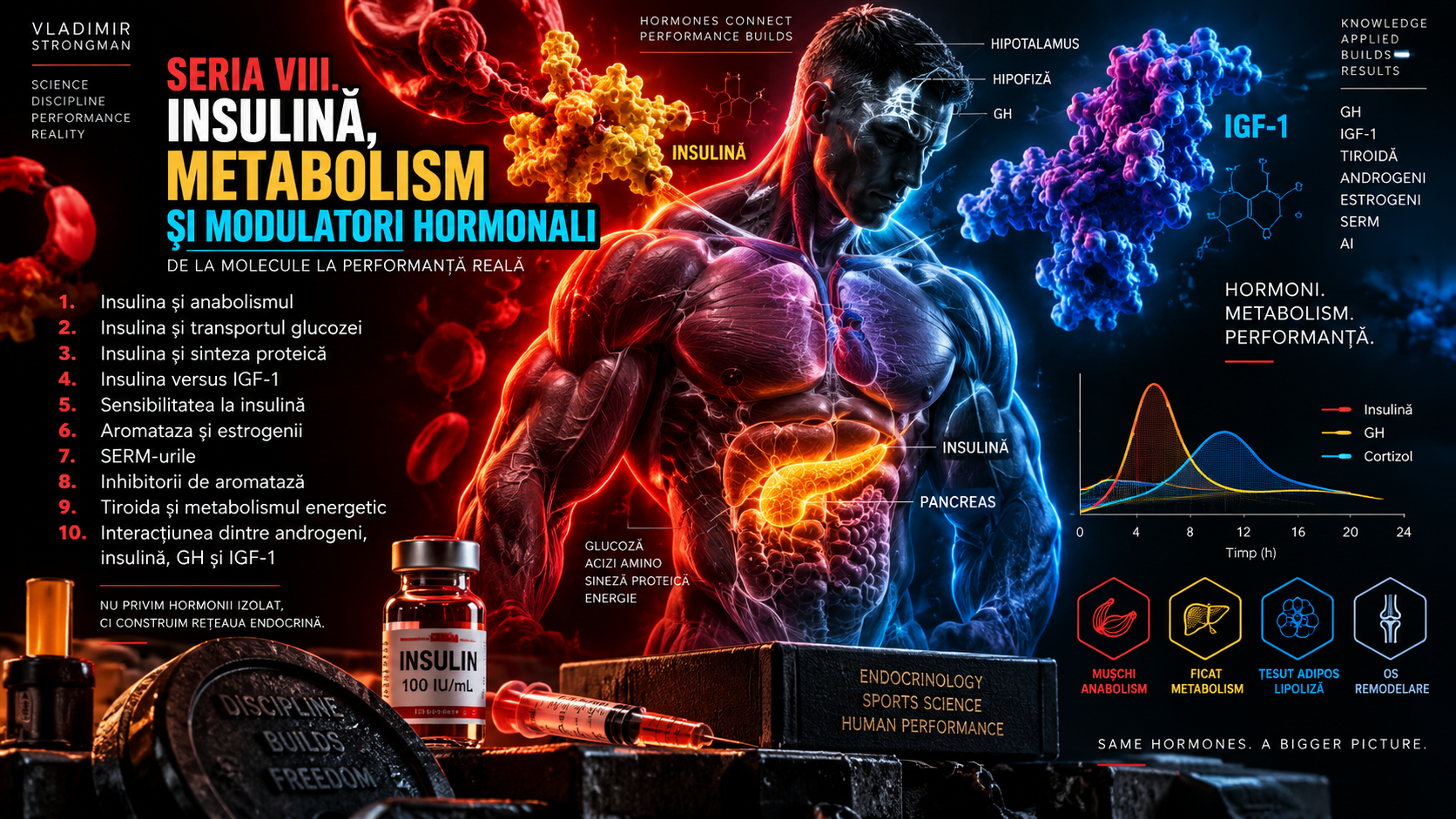
Insulin and IGF-1 are two molecules that often appear in the same conversation about metabolism, growth and performance. The name insulin-like growth factor 1 itself signals the relationship: IGF-1 is a growth factor with structural and functional similarities to insulin. This creates a persistent confusion. If the two molecules use similar signaling pathways and can activate Akt and mTOR, are they simply different versions of the same hormone? The answer is no.
In normal physiology, insulin is primarily a metabolic regulator. It responds rapidly to nutrient availability, suppresses hepatic glucose production, facilitates glucose uptake in muscle and adipose tissue, promotes glycogen storage and reduces lipolysis and proteolysis. IGF-1 operates with a different logic. It is integrated into the growth hormone–liver–IGF axis and acts as a mediator of growth, cell survival, skeletal and muscular development and tissue remodeling.
The two systems intersect in many places, but they should not be superimposed. This episode follows the boundary between them: what insulin and IGF-1 share, where they diverge, why their receptors are close relatives, how GH, liver and IGF-binding proteins enter the story, what happens in skeletal muscle and why an IGF-1 blood test cannot be interpreted as a simple measurement of “anabolism”.
1. Why is it called insulin-like growth factor?
IGF-1 is a peptide hormone structurally related to proinsulin. Evolutionarily, insulin, IGF-1 and IGF-2 belong to the same molecular family. Structural similarity also explains the relationship between their receptors. But evolution did not preserve two redundant copies of the same system. Insulin became specialized for rapid metabolic control, whereas the IGF system remained more strongly linked to growth, development and cell survival.
The term insulin-like does not mean that IGF-1 is simply a more anabolic insulin. It means there is molecular and functional homology. IGF-1 can produce some insulin-like effects, particularly at sufficiently high concentrations, and insulin can interact weakly with IGF-1R. Yet at physiological concentrations and in normal tissues, receptor specificity and receptor distribution preserve predominantly different functions.
2. Insulin: a hormone of the fed state
Insulin changes rapidly from meal to meal. The pancreatic beta cell tracks glucose and other nutrient availability, and secretion can rise within minutes. Temporally, insulin is a highly dynamic system. The body can move from basal fasting concentrations to postprandial elevations and then back toward baseline within hours.
This dynamic is perfectly suited to its role. After a meal, the body must limit the rise in blood glucose and decide where nutrients will go. Muscle takes up glucose, liver changes its metabolic fluxes, adipocytes reduce fatty-acid release and proteolysis declines. Insulin does not tell the body “grow for the long term”; it more closely communicates “nutrients are available now, use them and conserve them.”
3. IGF-1: a signal of growth and tissue state
Circulating IGF-1 is produced largely by the liver under the influence of growth hormone, but many tissues can produce IGF-1 locally. Muscle, bone and other organs can use IGF signaling in an autocrine or paracrine mode, meaning the molecule may act close to where it was produced. This local dimension matters and explains why the plasma concentration does not describe the entire IGF system.
IGF-1 varies more slowly than insulin and is influenced by age, nutritional state, GH secretion, liver function, sleep, illness and other factors. Concentrations are high during periods of growth and decline with age. Unlike insulin, which can respond abruptly to a meal, IGF-1 reflects a more integrated endocrine and nutritional state.
4. The insulin receptor and IGF-1R: two molecular cousins
The insulin receptor, IR, and the IGF-1 receptor, IGF-1R, belong to the same subfamily of receptor tyrosine kinases. Both are dimeric receptors with extracellular alpha subunits and beta subunits that span the membrane and contain the intracellular kinase domain. Ligand binding changes receptor conformation and activates autophosphorylation of tyrosine residues.
After this step, both IR and IGF-1R can recruit IRS proteins and activate PI3K, Akt and other cascades. This shared architecture creates the impression that the difference between insulin and IGF-1 is small. But biological outcome is not determined by one signaling diagram. Ligand-receptor affinity, receptor density, tissue, signal duration, adaptor proteins and the branches amplified in that context all matter.
5. The same Akt, different physiology
Both insulin and IGF-1 can activate Akt. In muscle, Akt can influence GLUT4, glycogen synthase, mTORC1 and FoxO factors. If we look only at this shared node, the differences seem to disappear. Physiology, however, works through distribution and dosing. Insulin signaling in muscle is strongly configured for carbohydrate and protein metabolism around meals, whereas IGF-1R signaling is more strongly configured toward growth and cell survival.
GLUT4 provides a useful example. Insulin is a major regulator of GLUT4 translocation and postprandial glucose uptake. IGF-1 can produce metabolic effects under some conditions, but it is not the principal hormone organizing daily glucose uptake after meals. IGF-1 instead has a more pronounced role in processes such as proliferation, differentiation, bone growth and tissue adaptation.
6. IR/IGF-1R hybrid receptors
The story becomes even more interesting because IR and IGF-1R can combine into hybrid receptors. Such a receptor contains one insulin-receptor half and one IGF-1-receptor half. They occur in tissues co-expressing both receptor types and generally display much higher affinity for IGF-1 than for insulin.
Hybrid receptors show that the separation between the insulin and IGF systems is not a wall. It is a permeable biological frontier. Their existence still does not mean that every IGF-1 effect can be reproduced by insulin or vice versa. Specificity is the product of the entire system, not merely the theoretical ability to activate a particular kinase.
7. The GH–liver–IGF-1 axis
A large fraction of circulating IGF-1 is linked to growth hormone. GH is secreted in pulses by the pituitary and acts on the GH receptor in liver and other tissues. GH-receptor activation primarily uses JAK2 and STAT5 to modify gene expression, including hepatic synthesis of IGF-1.
The classic model describes GH as the hormone transmitting the growth command and IGF-1 as a peripheral mediator. Reality is more complex. GH also has IGF-independent effects, while IGF-1 can be produced locally. Circulating IGF-1 additionally participates in negative feedback on the hypothalamic-pituitary axis and contributes to regulation of GH secretion.
8. IGFBPs: why IGF-1 does not circulate freely like insulin
One of the largest differences between insulin and IGF-1 is how they circulate in blood. Insulin is secreted and circulates largely as free hormone, with a half-life measured in minutes. IGF-1, by contrast, is overwhelmingly bound to transport proteins called insulin-like growth factor binding proteins, or IGFBPs.
IGFBP-3 is the most abundant circulating binding protein and forms, together with IGF-1 and the acid-labile subunit, a large ternary complex. This complex dramatically extends the lifetime of IGF-1 in circulation and limits its free passage through capillaries. The system behaves as a buffered reservoir rather than a hormone rising and falling sharply after every meal.
9. Total IGF-1 versus free IGF-1
When a laboratory measures IGF-1, it usually reports total IGF-1. Only a small fraction circulates freely or loosely bound and is immediately available to receptors. This distinction complicates biological interpretation. Two people can have similar total IGF-1 yet differ in how much is distributed among binding complexes and bioactive fractions.
Total IGF-1 nevertheless remains the most frequently used clinical marker because it is far more stable than GH, which is secreted in pulses. The value must be interpreted against age and the laboratory reference range. A single result does not tell us how active mTOR is inside muscle or how much muscle a person can build.
10. IGFBP-1 and the surprising connection to insulin
IGFBP-1 offers an elegant example of interaction between the two systems. Hepatic production of IGFBP-1 is inhibited by insulin. During fasting, when insulin is low, IGFBP-1 tends to rise and can reduce immediate IGF-1 availability. After a meal, rising insulin suppresses IGFBP-1 and thereby modifies the distribution of bioactive IGF-1.
This relationship shows that insulin and IGF-1 are not independent systems. Nutritional state influences the IGF axis partly through binding proteins. The body aligns energy availability with growth signaling. Biologically it would make little sense to maintain maximal growth programs during severe energy deprivation.
11. Nutrition and IGF-1
IGF-1 is sensitive to nutritional state. Severe energy restriction, malnutrition and inadequate protein intake can reduce IGF-1 even when GH secretion is normal or elevated. This phenomenon is sometimes described as hepatic GH resistance: the GH signal exists, but the liver no longer produces the same amount of IGF-1.
In an energy surplus the situation is not simply reversed. A higher IGF-1 value does not automatically mean greater hypertrophy. Muscle growth still requires training, amino acids, energy, recovery and mechanical adaptation. IGF-1 is a participant in that orchestration, not a single score of anabolic potential.
12. Locally produced IGF-1 in skeletal muscle
Skeletal muscle can produce IGF-1 locally, and this production can change with mechanical loading and tissue state. Autocrine and paracrine signaling allow adaptation to be concentrated in a loaded tissue without requiring the body to depend exclusively on circulating IGF-1.
This distinction matters when interpreting studies. An athlete can develop substantial muscular adaptation without a dramatic rise in serum IGF-1. Training can modify local IGF-system expression, receptor sensitivity and interactions with mechanotransduction, all of which are partly invisible in a routine blood test.
13. MGF: an isoform concept, not a separate magical hormone
Sports literature frequently uses the term MGF, mechano growth factor. It refers to transcript variants associated with the IGF-1 gene and mechanical response, not to the existence of a completely independent second IGF system. IGF-1 isoform biology is complex, and extrapolation from gene expression to commercial products promising hypertrophy is far stronger online than in human evidence.
It is important to separate local expression of IGF-1 variants after loading from the idea that a peptide carrying the MGF label would automatically reproduce the entire physiological response. Gene expression, precursor processing, local distribution and receptor interaction are all parts of a system that cannot be compressed into one commercial name.
14. IGF-1 and mTORC1
IGF-1R can activate PI3K and Akt, and Akt can facilitate mTORC1 activation by inhibiting TSC1/TSC2 and modifying PRAS40. Through mTORC1, the signal can influence p70S6K1, 4E-BP1, translation initiation and protein-synthetic capacity. This is one molecular basis for the role of IGF-1 in muscle growth.
Insulin can activate the same region of the network. The difference is not that IGF-1 possesses mTOR while insulin does not. The difference is how, where and for how long the network is activated and what other signals are present. Amino acids, mechanical tension and energy state remain essential. mTORC1 is an integrator, not the private property of one hormone.
15. FoxO: a point where both systems can reduce catabolism
Akt can phosphorylate FoxO transcription factors and prevent them from activating nuclear programs associated with atrophy. Through this route, both insulin and IGF-1 can reduce expression of E3 ligases such as MuRF1 and atrogin-1 in some experimental contexts.
Again, molecular overlap does not mean physiological identity. Insulin normally provides an anti-catabolic brake closely linked to nutrient availability. IGF-1 participates in a broader program of tissue maintenance, growth and regeneration. The same FoxO node can be reached from two endocrine systems with different missions.
16. IGF-1 and satellite cells
Satellite cells are muscle stem cells located between the fiber membrane and basal lamina. They can be activated after injury or loading and can proliferate, differentiate and fuse with existing fibers. The IGF system participates in regulation of several stages of this process, from proliferation through differentiation.
This role helps explain the association between IGF-1 and regenerative capacity. Insulin can support the metabolic environment needed for repair, but it is not usually described as the central regulator of satellite-cell proliferation and differentiation. Here the difference between metabolism and growth becomes more visible.
17. Bone and cartilage: where IGF-1 clearly separates from insulin
IGF-1 is essential for skeletal development and growth-plate function. It acts on chondrocytes and osteoblasts and mediates an important part of GH effects on longitudinal growth. In adults, the IGF system continues to participate in bone remodeling.
Insulin also influences bone directly and indirectly, but it cannot physiologically replace the GH–IGF axis in skeletal development. This is a clear demonstration that two molecules capable of activating PI3K-Akt are not automatically equivalent at the level of the whole organism.
18. Insulin and glucose versus IGF-1 and growth
If the difference must be reduced to one simple image, insulin primarily manages substrate flow, whereas IGF-1 primarily manages programs of growth and adaptation. Insulin rapidly determines whether glucose is taken up and whether lipolysis is suppressed. IGF-1 changes, over a longer time scale, the probability that a cell will grow, survive or differentiate.
This simplification is not perfect. Insulin is anabolic and IGF-1 has metabolic effects. But as a conceptual map it is much more accurate than the idea that IGF-1 is simply a stronger insulin.
19. Protein synthesis: where IGF-1 has a clearer growth-factor profile
In muscle, IGF-1 can increase anabolic signaling through PI3K-Akt-mTOR and reduce some catabolic pathways through FoxO. These mechanisms are well demonstrated experimentally. Yet the transition from molecular signaling to long-term human hypertrophy is not a straight line. Mechanical training remains the main stimulus deciding which tissue must adapt.
Insulin also has anabolic effects on protein metabolism, but in humans its most robust effect is anti-catabolic: it reduces protein breakdown and improves net protein balance when amino acids are available. Therefore, if the question is “which is more oriented toward cellular growth?”, the answer is IGF-1. If the question is “which organizes daily nutrient metabolism?”, the answer is insulin.
20. Insulin controls blood glucose much more directly
Insulin is central to postprandial glucose control. It increases glucose uptake in muscle and adipocytes through GLUT4 and suppresses hepatic glucose production. Severe insulin deficiency, as in untreated type 1 diabetes, produces hyperglycemia, uncontrolled lipolysis and ketogenesis. IGF-1 cannot physiologically take over this function.
IGF-1 can have glucose-lowering effects because it can activate members of the insulin-receptor family and increase glucose use. But this is secondary to its main role and can become clinically relevant through hypoglycemia risk when the system is pharmacologically manipulated.
21. GH can move metabolically in the opposite direction from insulin
Growth hormone is frequently associated with IGF-1, but metabolically the two are not identical. GH stimulates lipolysis and can reduce insulin sensitivity, increasing fatty-acid availability and helping preserve circulating glucose. In chronic excess, this action can promote hyperinsulinemia and impaired glucose tolerance.
IGF-1, by contrast, has some insulin-like effects and can improve glucose utilization under certain conditions. The GH–IGF axis therefore contains two components that cooperate in growth but are not metabolic twins. One reason endocrinology is so complex is that one axis can contain seemingly opposite signals for carbohydrate metabolism.
22. Hyperinsulinemia does not automatically mean high IGF-1
In insulin resistance, the pancreas may secrete more insulin to maintain glucose control. This hyperinsulinemia can influence IGFBP-1 and other components of the IGF axis, but there is no simple conversion between insulin and IGF-1. Liver, GH, nutritional status, inflammation and body composition modify the system independently.
This is why fasting insulin and IGF-1 answer different questions. Insulin can provide information about metabolic regulation and, together with glucose, may enter estimates such as HOMA-IR. IGF-1 is used clinically mainly to evaluate the GH axis. Combining them into a single “anabolism score” has no physiological basis.
23. The liver is central to both systems in different ways
The liver receives insulin at high concentration immediately after pancreatic secretion because pancreatic blood drains into the portal circulation. There insulin suppresses gluconeogenesis and glycogenolysis, promotes glycogen synthesis and changes lipid metabolism.
The same liver produces most endocrine IGF-1 under GH influence and also synthesizes multiple IGFBPs and the acid-labile subunit. Thus the liver is simultaneously a major insulin target and the main factory for circulating IGF-1. This explains why liver disease can profoundly alter both glucose metabolism and IGF-1 values.
24. Resistance training does not require a large rise in serum IGF-1 to produce hypertrophy
One important lesson from training physiology is that circulating hormones should not be confused with local muscle signals. Hypertrophy after resistance training can occur without spectacular chronic increases in serum IGF-1. Muscle responds to mechanical tension, stretch, recruitment, amino acids and energy through a network of mechanosensors and local signals.
This matters for athletes who view hormone tests as a ranking of progress. An IGF-1 value near the upper reference range does not guarantee a superior hypertrophic response, and an average value does not mean muscle is unable to grow. Adaptation cannot be compressed into one biomarker.
25. Mechanotransduction can activate growth independently of systemic IGF-1
Modern models of hypertrophy show that mechanical tension can activate mTORC1 and related pathways through mechanisms that do not obligatorily require systemic IGF-1 signaling. Integrins, focal-adhesion complexes, phosphatidic acid and other mechanosensitive systems can convert physical force into biochemical information.
This does not make IGF-1 irrelevant. It means muscle has several inputs into the same anabolic infrastructure. Biological redundancy is an advantage: an organism does not base its entire adaptation to loading on one endocrine molecule.
26. IGF-1 and regeneration after injury
After muscle injury, regeneration requires controlled inflammation, removal of damaged tissue, satellite-cell activation, proliferation, differentiation, revascularization and remodeling of extracellular matrix. The IGF system participates in several stages of this sequence.
Insulin contributes indirectly by supporting an energetic and anti-catabolic environment, but its role is not identical. This is why the word “anabolic” can hide major differences: protein conservation and regenerative stimulation are both favorable to tissue, yet they are distinct biological processes.
27. IGF-1 and age
IGF-1 follows a strongly age-dependent curve. Levels rise during childhood, reach high values during puberty and adolescence, and then progressively decline through adulthood. Reference intervals therefore need to be age-adjusted. A value normal for an adult could be too low for an adolescent and vice versa.
Insulin does not follow the same developmental curve. It is regulated moment to moment by glucose, insulin sensitivity and metabolic demand. Puberty can temporarily alter insulin sensitivity, but the temporal logic of the system remains different from IGF-1.
28. Sleep, GH and IGF-1
GH secretion is closely linked to sleep, with important pulses associated with deep sleep. Sleep deprivation and circadian disruption can alter the GH axis and metabolism. IGF-1, being more stable, does not reproduce each GH pulse but reflects longer-term function of the axis.
Insulin is also affected by sleep, but through a different dominant mechanism: sleep restriction can reduce insulin sensitivity and impair glucose control. Thus the same poor night can influence both systems, but not in the same way.
29. Energy deficit: why both systems change
In an energy deficit, insulin tends to be lower because nutrient intake and glucose availability decline, while lipolysis becomes more permissive. If the deficit is moderate and the person remains active, insulin sensitivity may improve.
During severe or prolonged restriction, IGF-1 can fall, reflecting the body's prioritization of survival and energy conservation over growth. This response again shows that IGF-1 belongs to a system asking not only “are nutrients available now?” but also “are there enough resources to sustain growth?”
30. Obesity and insulin resistance change the terrain
In obesity and insulin resistance, the relationships among insulin, GH and IGF-1 can shift in complex ways. Hyperinsulinemia can suppress IGFBP-1, GH secretion may be reduced, and total IGF-1 may sometimes remain within range. There is no universal pattern that can be reduced to simply “high IGF-1” or “low IGF-1.”
This is an important reason biomarkers must be interpreted in context. Waist circumference, body composition, glucose, insulin, lipids, liver function, sleep and activity level can change the meaning of the same endocrine value.
31. High IGF-1 does not automatically mean “more anabolic”
It is tempting to view IGF-1 as an anabolism dial: low means little and high means a lot. Biology does not work that way. Serum IGF-1 is influenced by age, nutrition, GH, liver and binding proteins. The final effect also depends on receptor availability, tissue context and local signals.
A high value can be physiological during adolescence, can appear with excessive GH-axis activity or can reflect other endocrine characteristics. It is not a “muscle-mass score.” Likewise, high insulin does not automatically mean superior anabolism; in insulin resistance it may indicate that the body requires more hormone to obtain the same metabolic effect.
32. Low IGF-1 does not automatically mean growth-hormone deficiency
Low IGF-1 can occur in GH deficiency, but also in malnutrition, liver disease, systemic inflammation, severe hypothyroidism and other catabolic states. Endocrine diagnosis therefore cannot be made from one number. Clinicians interpret symptoms, history, physical findings and, when needed, dynamic testing of the GH axis.
Insulin teaches the same methodological lesson. Low fasting insulin can be excellent in an insulin-sensitive person and problematic in pancreatic insufficiency. A biomarker makes sense only in relation to the clinical question.
33. GH and IGF-1 excess: the lesson of acromegaly
Acromegaly provides a natural example of what happens when the GH–IGF axis is excessively active for a long time. Patients may develop soft-tissue enlargement, skeletal changes, organ enlargement, sleep apnea, hypertension and cardiovascular problems. Metabolically, excessive GH can promote insulin resistance and diabetes.
This picture is an important antidote to the idea that “more growth factor” is synonymous with “more healthy performance.” Growth signaling is not selective for the biceps or quadriceps. Receptors are distributed throughout the body.
34. Severe IGF-1 deficiency: what physiology teaches us
Severe IGF-1 deficiency or GH resistance during childhood can produce major growth disorders. These conditions show that IGF-1 is essential for normal development, not merely an anabolic bonus. Its role is deeply integrated into skeletal, organ and tissue biology.
Severe insulin deficiency is a completely different metabolic emergency. Without sufficient insulin, glucose is no longer handled appropriately, lipolysis and ketogenesis become uncontrolled and life can be threatened rapidly. Comparing the two deficiencies again highlights their primary roles: acute metabolism versus growth and development.
35. Mecasermin: IGF-1 as a medicine
A recombinant form of IGF-1, mecasermin, is used medically in certain rare forms of severe primary IGF-1 deficiency. The existence of an approved medicine does not make the molecule a general recovery or hypertrophy agent for healthy people. Medical indication, patient selection and monitoring are essential.
Pharmacological IGF-1 can cause adverse effects including hypoglycemia, and its growth effects are not restricted to muscle. Medical use therefore occurs within a controlled framework. This article does not provide doses, combinations or non-medical use schedules.
36. Why IGF-1 can cause hypoglycemia
IGF-1 can increase glucose uptake and reduce hepatic glucose production through insulin-like effects. At pharmacological concentrations, this component can become strong enough to lower blood glucose. The risk demonstrates that overlap with the insulin system is biologically real rather than merely a textbook similarity.
The fact that both can cause hypoglycemia still does not make them equivalent. Insulin is the dominant physiological regulator of blood glucose, whereas IGF-1's metabolic effect is one component of a broader growth-factor profile.
37. Why the receptor matters more than the molecule's label
A molecule produces effects through the receptors it activates and the context in which those receptors exist. Insulin has very high affinity for IR, IGF-1 has very high affinity for IGF-1R, and hybrid receptors generally favor IGF-1. At pharmacological concentrations, specificity can blur and cross-reactivity becomes more important.
This rule appears throughout endocrine pharmacology: an effect is not defined by the label “anabolic,” but by pharmacokinetics, concentration, receptor, tissue and duration. When concentrations move far outside the physiological range, effects minor or absent under normal conditions can become relevant.
38. Insulin versus IGF-1 in muscle: the short comparison
Insulin facilitates glucose uptake, supports glycogen synthesis, reduces protein breakdown and coordinates the postprandial state. IGF-1 signals growth and survival and contributes to protein synthesis, regeneration, differentiation and satellite-cell biology. Both can activate Akt and mTORC1, but the physiological reasons for activating the network differ.
A useful analogy is a construction site. Insulin largely handles logistics: fuel, available materials, reducing losses and storing resources. IGF-1 resembles a development and expansion program. Yet neither logistics nor expansion builds useful structure without the mechanical blueprint supplied by training and the raw material supplied by amino acids.
39. What this difference means for Strongman
Strongman places large demands on metabolism and on structures that must adapt. Carries, medleys and repetition events consume glycogen and place obvious demands on glucose regulation. Here the role of insulin and insulin sensitivity is evident in restoration of substrates between sessions.
At the same time, muscle mass, connective tissue and bone must tolerate years of extreme loading. The GH–IGF axis participates in the biology of these tissues, but performance cannot be inferred from serum IGF-1. A Strongman athlete with good programming, adequate protein and energy, sufficient sleep and mechanical progression can build adaptation without artificially chasing one hormone value.
40. Laboratory testing: why IGF-1 and fasting insulin are not read the same way
IGF-1 is relatively stable across the day compared with GH and is usually interpreted with age-dependent reference ranges. Fasting insulin is much more closely tied to immediate metabolic state and must be interpreted alongside glucose, body composition, diet and other information.
Neither test is a direct hypertrophy assay. A laboratory can provide information about an endocrine axis or metabolism, but it cannot replace measurement of performance, body composition and training progress.
41. The myth that “IGF-1 is insulin for muscle”
This phrase is attractive because it compresses two complex systems into one sentence. But it misses exactly what matters: different receptors, IGFBPs, the GH axis, local production, roles in development and bone, satellite-cell effects and signal duration.
A more accurate statement is that insulin and IGF-1 belong to the same molecular family and share important signaling infrastructure, while that infrastructure is used for different physiological objectives.
42. Anti-doping: two systems, two categories
On the WADA 2026 Prohibited List, IGF-1 and its analogues are included among prohibited growth factors in category S2, while insulins and insulin-mimetics are listed under S4, Hormone and Metabolic Modulators. Both categories are prohibited at all times, in and out of competition, unless a valid therapeutic-use framework applies.
This anti-doping separation mirrors the physiological distinction discussed throughout the article: IGF-1 is treated as a growth factor, whereas insulin is treated as a metabolic modulator. The classification is not a complete endocrinology lesson, but it is remarkably intuitive.
43. What should you remember?
Insulin and IGF-1 are relatives, not twins. Their molecules are related, their receptors are related and their signaling pathways overlap, but their principal functions differ. Insulin is the rapid regulator of nutrient availability and storage. IGF-1 is a central mediator of growth, development and remodeling.
In muscle, both contribute to an anabolic environment, yet neither replaces mechanical tension, amino acids, energy and time. Insulin can reduce breakdown and support substrate uptake; IGF-1 can activate programs of growth and regeneration. Real hypertrophy is the product of integrating all these signals.
In laboratory testing, insulin and IGF-1 should be interpreted for different questions. High IGF-1 is not a certificate of anabolism, and high insulin is not an advantage when it reflects insulin resistance. Context turns a number into information.
44. PI3K-Akt is not the only pathway: MAPK-ERK
Both the insulin receptor and IGF-1R can activate, in addition to PI3K-Akt, the Ras-Raf-MEK-ERK family. This branch is more closely associated with changes in gene expression, proliferation and differentiation. IGF-1R is generally viewed as more strongly oriented toward these mitogenic effects, whereas the insulin receptor is more strongly associated with rapid metabolic responses.
The separation is not absolute. Insulin can activate ERK and IGF-1 can activate metabolic pathways. The proportion between branches and the duration of signaling help create biological identity. A short, meal-associated metabolic activation of Akt is a different problem from a signaling program modifying proliferation and differentiation over longer periods.
45. Insulin receptor isoforms: IR-A and IR-B
The insulin receptor itself is not one perfectly uniform entity. Alternative splicing produces two major isoforms, IR-A and IR-B. IR-B is more strongly associated with classical metabolic functions and is abundant in tissues such as liver, whereas IR-A has a more mitogenic profile and can bind IGF-2 with greater affinity.
This distinction adds another layer to the insulin-IGF comparison. Not only the ligand matters, but also the receptor version present in a tissue. Two cells can receive the same molecule and produce different responses because they express receptors, adaptor proteins and enzymes in different proportions.
46. Where does IGF-2 fit?
IGF-2 is the third major member of the insulin-IGF family. It plays a major role in fetal development and can signal through IGF-1R and IR-A. Its existence shows that the family is not organized as a duel between two molecules but as a network of ligands and receptors with overlapping affinities.
IGF-2 also binds the IGF-2/mannose-6-phosphate receptor, which functions largely in ligand uptake and degradation rather than as a classical receptor tyrosine kinase like IGF-1R. This mechanism helps control growth-factor availability and demonstrates how tightly regulated the family is.
47. Receptors do not stay permanently active: internalization and feedback
After activation, insulin receptors and IGF-1R can be internalized, recycled or degraded. In parallel, cells activate feedback mechanisms that reduce signal intensity. Phosphatases remove phosphate groups, while feedback proteins can modify IRS and other network components.
This explains why hormonal signaling cannot be understood by the rule “more ligand always produces proportionally more effect.” Receptors can saturate, signaling can desensitize and intracellular feedback can limit the response. Endocrinology is built on dose-response curves and control systems, not infinity.
48. Acute hormonal increases after training do not explain hypertrophy by themselves
Hard training sessions can transiently change GH, insulin and other circulating signals. In the past, these acute spikes were sometimes treated as a primary explanation for hypertrophy. Modern research has greatly weakened that idea. Muscle adaptation is more directly connected to mechanical tension, effective volume, recruitment, nutrition and local signaling than to the size of a systemic hormone spike lasting a few dozen minutes.
This observation matters for insulin versus IGF-1. The important question is not which one “rises more” after a session but what function each system serves over the hours and days of recovery. A hormone can be important without the amplitude of its acute spike predicting progress.
49. IGF-1, tendons and connective tissue
The IGF system is also involved in fibroblast biology, extracellular-matrix synthesis and connective-tissue responses. This has generated interest in IGF-1 in tendon biology and repair. Tendon adaptation, however, is slow and depends on mechanical loading, collagen organization, vascular factors and numerous local signals.
There is no basis for assuming that one growth factor can turn a tendon into a resilient structure without appropriate mechanical progression. More signaling does not automatically mean better collagen architecture. For Strongman this nuance matters because muscle strength can increase faster than the capacity of some connective structures to tolerate load.
50. Growth is not selective for muscle alone
IGF-1R is expressed in many tissues. A systemic growth signal does not carry a map labeled “skeletal muscle only.” Bone, cartilage, organs and other cell types also respond to the GH-IGF axis. Extrapolating an anabolic pathway into the idea of selective muscle hypertrophy is therefore biologically naive.
Insulin is equally non-selective from another perspective. Insulin receptors exist in liver, adipocytes, muscle, brain and many other tissues. A systemic metabolic signal distributes effects according to tissue and physiological state. No major circulating hormone is designed exclusively for sports performance.
51. Laboratory variability: why a trend can matter more than one blood draw
IGF-1 is more stable than GH but is not immune to biological and analytical variation. Nutritional state, recent illness, liver function and assay methodology can influence the result. A surprising value is therefore often interpreted through repetition and clinical context rather than immediate dramatic conclusions.
Fasting insulin can vary even more with sleep, previous dietary intake, stress and metabolic sensitivity. For monitoring, similar collection conditions and change over time are more informative than obsessively comparing two numbers obtained under different circumstances.
52. Insulin versus IGF-1 has no universal winner
The question “which is more anabolic?” is too vague to have a good answer. More anabolic for what? For conserving protein after a meal, insulin is crucial. For longitudinal growth, IGF-1 is indispensable. For glucose uptake, insulin dominates. For programs of proliferation and differentiation, IGF-1R plays the more pronounced role.
In the real organism the two do not compete. They cooperate within a network integrating nutrients, energy, GH, amino acids and mechanical loading. Insulin communicates that resources are available. IGF-1 contributes to growth and remodeling programs. Muscle determines how it responds according to what has been mechanically demanded of it.
Viewed together, the two systems illustrate a fundamental biological rule: growth is permitted only when metabolism can support it. Insulin connects immediate nutrient availability with use and storage, whereas IGF-1 connects energy state and the GH axis with programs of tissue development. Their overlap is not a design flaw. It is part of the mechanism by which the body prevents growth from becoming disconnected from the resources required to sustain it.
Main sources
Siddle K. Signalling by insulin and IGF receptors: supporting acts and new players. Journal of Molecular Endocrinology. 2011.
Simmons KJ, Fishwick CWG. Recent developments in the structural characterisation of the insulin receptor and IGF1R, including hybrid receptors. 2022.
Yoshida T, Delafontaine P. Mechanisms of IGF-1-mediated regulation of skeletal muscle hypertrophy and atrophy. Cells. 2020.
Philippou A, Maridaki M, Halapas A, Koutsilieris M. The role of the IGF-1 system in skeletal muscle. In Vivo. 2007.
Clemmons DR. Modifying IGF1 activity: an approach to treat endocrine disorders, atherosclerosis and cancer. Nature Reviews Drug Discovery. 2007.
Glass DJ. Skeletal muscle hypertrophy and atrophy signaling pathways. International Journal of Biochemistry & Cell Biology. 2005.
Bodine SC et al. Akt/mTOR pathway in skeletal muscle hypertrophy and atrophy. Nature Cell Biology. 2001.
World Anti-Doping Agency. The 2026 Prohibited List and explanatory notes.
Editorial note: this article is educational and does not constitute medical advice or a protocol for insulin, IGF-1, GH or other hormonal modulators. Pharmacological manipulation of these systems can cause hypoglycemia, metabolic disturbances and other serious complications.
More From The Journal

Training
The Science of Strongman Training - Episode X: Competition Programming
How to build a complete Strongman competition preparation block: calendar, events, volume, intensity, specificity, simulations, autoregulation, tapering and fatigue management.

Training
The Science of Strongman Training - Episode IX: Peaking
Peaking in Strongman: how to turn months of training into maximal competition performance by reducing fatigue, preserving adaptations and managing specificity.