
Pharmacology
Series: Insulin, Metabolism and Hormonal Modulators - Episode 2: Insulin and Glucose Transport
September 8, 2026
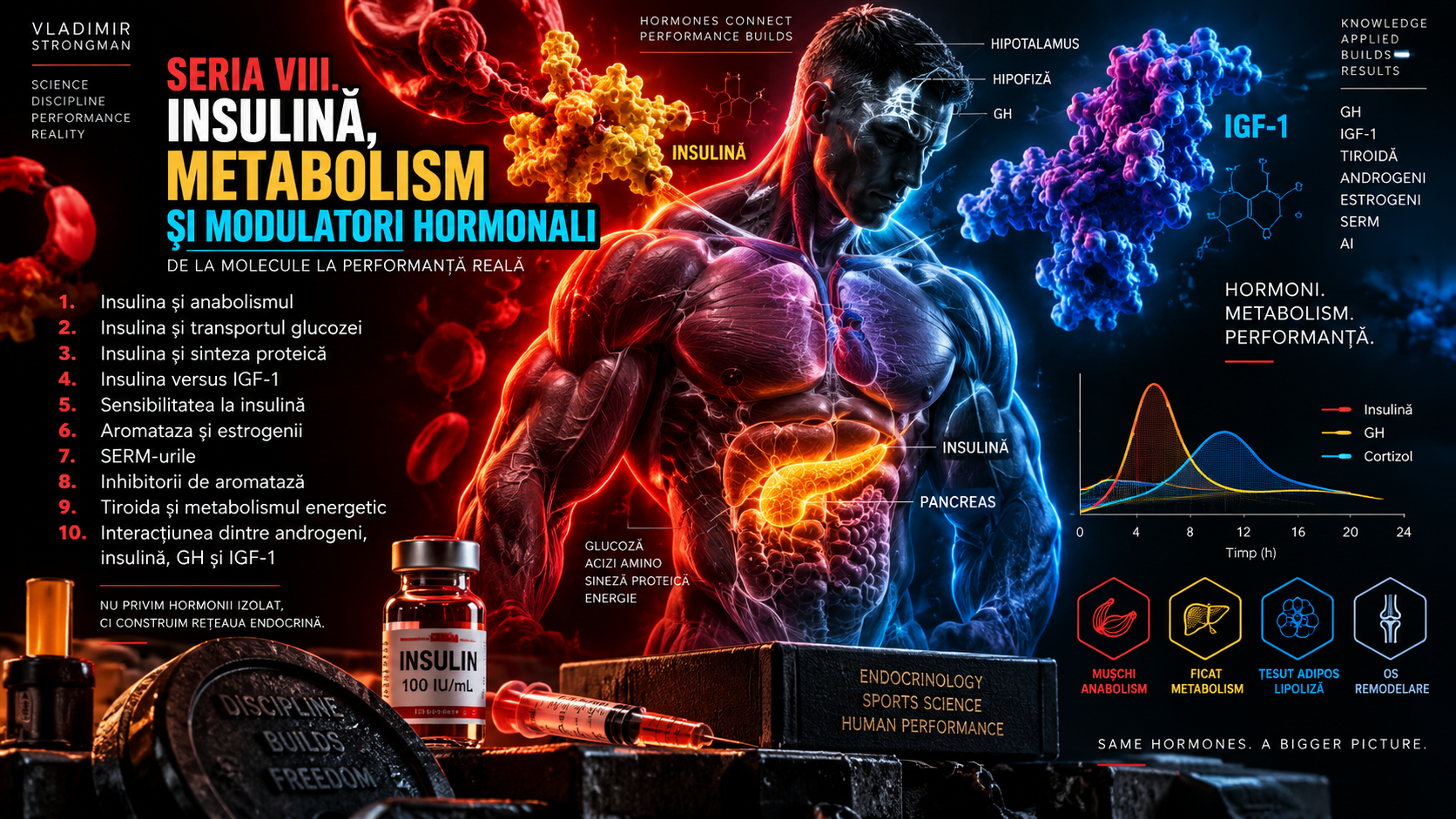
In the previous episode we established that insulin is more than a blood-glucose hormone. Episode 2 moves into the mechanism at the center of that story: how glucose travels from the extracellular space into cells and how insulin changes the speed of that process. The question sounds simple, but the answer involves membrane transporters, enzymes, intracellular vesicles, kinase signaling, blood flow, muscle contraction and the energetic state of the cell.
One of the most persistent misconceptions is that insulin “opens the cell” and pushes glucose inside. In reality, glucose crosses membranes through specialized transport proteins, while insulin changes the availability and activity of some of those transporters in specific tissues. Skeletal muscle is the classic example because the GLUT4 transporter can be moved rapidly from intracellular compartments to the cell surface.
For an athlete, this subject is fundamental. Glucose and glycogen support a major part of repeated high-intensity work, and the muscle's capacity to take up and store glucose influences recovery between sessions. The same mechanisms also matter for health: insulin resistance, type 2 diabetes and ectopic fat accumulation alter how these pathways respond to nutrients.
1. Glucose: circulating fuel and metabolic raw material
Glucose is a six-carbon monosaccharide and one of the main forms in which energy derived from carbohydrate circulates in blood. After intestinal absorption, some is used immediately, some is stored as glycogen, and under certain conditions some can contribute to lipid synthesis. Glucose matters not only because it can generate ATP but also because it supplies carbon to many biosynthetic pathways.
The cell membrane is largely a lipid bilayer, while glucose is a polar molecule. It cannot cross this barrier rapidly by simple diffusion. The body therefore uses transport proteins. Some allow facilitated diffusion down a concentration gradient, while others indirectly use the energy stored in an ion gradient to move glucose. Without these systems, blood-glucose control and tissue fueling would be impossible.
2. The GLUT family: there is no single glucose transporter
GLUT transporters belong to the SLC2 family and are not interchangeable. Differences in tissue expression, substrate affinity, capacity and regulation allow each organ to handle glucose according to its function. GLUT1, GLUT2, GLUT3 and GLUT4 are especially useful for understanding human metabolism, although the family includes additional members and some transport other hexoses.
Saying that “insulin transports glucose into cells” hides this diversity. Brain, liver, red blood cells and skeletal muscle do not use the same strategy. Some tissues take up glucose largely independently of insulin, whereas skeletal muscle and adipose tissue rely heavily on insulin-regulated GLUT4 trafficking. This difference becomes crucial when discussing hypoglycemia and insulin resistance.
3. GLUT1: basal transport that keeps cells supplied
GLUT1 is expressed in many tissues and is important for basal glucose uptake. Red blood cells use it extensively, and barriers such as the cerebral endothelium depend on it to maintain continuous glucose delivery to the nervous system. GLUT1 has relatively high affinity for glucose, allowing efficient transport even when plasma glucose is not especially high.
This basal transport is one reason severe hypoglycemia can become dangerous quickly. The brain needs a continuous substrate supply, and the fact that its transport does not primarily depend on GLUT4 does not protect it when circulating glucose becomes insufficient. The problem is not the absence of an insulin “key”, but lack of substrate available to glucose-dependent tissues.
4. GLUT2: a high-capacity transporter and metabolic gateway
GLUT2 is classically associated with liver, pancreatic beta cells, kidney and intestine, although exact distribution and function differ between species and cell types. It has high transport capacity and lower affinity for glucose than GLUT1 or GLUT3. That combination suits tissues that must respond across a broad range of glucose concentrations.
In hepatocytes, GLUT2 transport is bidirectional and is not regulated by insulin-dependent translocation in the way GLUT4 is. The liver can take up glucose after a meal and release glucose during fasting. Insulin strongly changes what the liver does with glucose after membrane transport by regulating glucokinase, glycogen synthesis and hepatic glucose production. The liver is therefore insulin-responsive even though glucose entry is not GLUT4-dependent.
5. GLUT3: high-affinity transport for nervous tissue
GLUT3 is expressed especially in neurons and has high affinity for glucose. This helps neurons capture glucose efficiently from brain extracellular fluid, where concentrations are lower than in plasma. The brain has high energy demand, and its transport system is designed for continuity rather than waiting for post-meal insulin peaks.
Insulin still has effects in the central nervous system on appetite, neuronal function and metabolism, but these should not be confused with the idea that neurons require insulin for basic access to glucose. This distinction matters because insulin-induced hypoglycemia becomes dangerous precisely when circulating glucose falls while the brain continues consuming substrate.
6. GLUT4: the transporter linking insulin to skeletal muscle
GLUT4 is the central transporter for insulin-stimulated glucose uptake in skeletal muscle and adipose tissue. In the absence of stimulation, much of GLUT4 is stored in specialized intracellular vesicles. When insulin signaling is activated, those vesicles move toward the membrane, fuse with it and rapidly increase the number of transporters available at the surface.
This mechanism produced the popular metaphor that insulin “opens the door”. The metaphor is useful at an introductory level but biologically incomplete. Insulin does not create a hole in the membrane. It triggers a signaling cascade that changes vesicle traffic and GLUT4 density at the cell surface. When the stimulus declines, part of the transporter pool is internalized again.
7. The insulin receptor: the start of the signal, not the transporter itself
The insulin receptor is a transmembrane protein with tyrosine-kinase activity. It contains extracellular subunits that bind insulin and membrane-spanning subunits that transmit the signal inward. Insulin binding changes receptor conformation and promotes autophosphorylation of tyrosine residues. These modifications create docking platforms for adaptor proteins, especially the IRS family, which carry the message downstream.
The receptor must be distinguished from the transporter. The insulin receptor detects the hormone and initiates signaling; GLUT4 is the protein that actually facilitates glucose movement across the membrane. A chain of events lies between them. A defect at the receptor, IRS proteins, intermediate enzymes or vesicle trafficking can reduce the final response even when GLUT4 protein is present in the cell.
8. IRS, PI3K and Akt: the main metabolic signaling axis
After receptor activation, IRS proteins are phosphorylated on tyrosine residues and can recruit phosphatidylinositol-3-kinase, or PI3K. PI3K modifies membrane lipids and generates signals that allow activation of PDK1 and Akt, also called protein kinase B. Akt is one of the central nodes in insulin action and influences glucose transport, glycogen synthesis, lipid metabolism and several processes related to cell growth.
For glucose transport, the key point is that Akt does not physically pull glucose into the cell. It phosphorylates targets that control GLUT4 vesicle traffic. At the same time, insulin signaling changes enzymes that determine what happens to glucose once it enters. Glucose uptake is therefore the coordinated product of membrane transport, phosphorylation and intracellular metabolism, not merely a surface-membrane event.
9. TBC1D4, Rab proteins and GLUT4 vesicle traffic
An important Akt target is TBC1D4, historically known as AS160. In the basal state, TBC1D4 helps keep certain Rab proteins in a state that limits movement of GLUT4 vesicles toward the membrane. Insulin-stimulated phosphorylation changes this molecular brake and allows Rab proteins to coordinate mobilization, targeting and movement of vesicles toward the cell surface.
This level of detail shows why the phrase “insulin activates GLUT4” is incomplete. The transporter does not simply run faster. A population of vesicles must leave intracellular compartments, travel along cytoskeletal structures, approach the membrane, dock and then fuse. Each step uses distinct proteins and can be influenced by the metabolic state of the cell.
10. Membrane fusion: SNARE proteins and the final meters of the route
When a GLUT4 vesicle reaches the plasma membrane, docking and fusion proteins become critical. SNARE complexes allow precise alignment of the two membranes and the fusion event that inserts GLUT4 into the cell surface. Proteins such as vesicular VAMP2 and membrane-associated syntaxin-4 and SNAP23 participate in this process together with many regulatory partners.
This biology matters because insulin sensitivity does not depend on one switch. A cell can contain a relatively normal amount of GLUT4 yet have a defect in proximal signaling or vesicle traffic that reduces translocation. Conversely, training can improve several links at once, increasing both GLUT4 abundance and the efficiency of the machinery that mobilizes it.
11. After glucose enters: hexokinase preserves the favorable gradient
Glucose entry is only the beginning. In muscle fibers, hexokinase rapidly phosphorylates glucose to glucose-6-phosphate. This reaction has two major consequences. First, glucose-6-phosphate cannot simply leave through GLUT4, so glucose is metabolically trapped inside the cell. Second, lowering free intracellular glucose helps maintain the concentration gradient favoring continued movement of glucose from blood toward muscle.
Glucose-6-phosphate sits at a metabolic intersection. It can enter glycolysis to produce ATP, contribute to glycogen synthesis, or enter the pentose-phosphate pathway and other processes. During intense exercise, glycolytic flux rises. After exercise, when immediate ATP demand falls and glycogen stores are reduced, a larger fraction can be directed toward storage restoration.
12. Insulin and glycogen synthesis
Glycogen is the polymeric storage form of glucose in liver and muscle. In muscle, the reserve is used locally for contraction and cannot be exported directly into blood as free glucose because skeletal muscle lacks meaningful glucose-6-phosphatase activity. Insulin promotes storage by increasing glucose uptake and by stimulating glycogen-synthase activity.
Part of this effect is mediated by Akt and inhibition of GSK3, a kinase that can phosphorylate and inhibit glycogen synthase. Glucose-6-phosphate also regulates the enzyme allosterically. Transport and storage are therefore coupled: more glucose entering and being phosphorylated provides both substrate and signaling support for glycogen restoration. After training, this coordination becomes especially efficient.
13. Blood flow and capillary recruitment: glucose must first reach the fiber
Glucose transport does not begin at the sarcolemma. Nutrients must first be delivered through the circulation to muscle capillaries, cross the endothelium and enter the interstitial space. Insulin can influence microcirculation through nitric-oxide-dependent endothelial signaling, while greater perfusion and capillary recruitment can increase the surface area through which insulin and glucose are delivered to muscle.
In insulin resistance, microvascular dysfunction may contribute to reduced hormone and substrate delivery. In athletes, exercise itself produces major changes in blood flow, and active muscle receives far more perfusion than resting muscle. Glucose uptake should therefore be viewed as a serial process: vascular delivery, membrane transport and intracellular metabolism.
14. Muscle contraction can move GLUT4 without a large insulin peak
One of the most important findings in exercise physiology is that contracting muscle can increase glucose uptake through mechanisms partly independent of insulin. When a fiber contracts, ATP turnover rises, AMP and ADP change, cytosolic calcium oscillates, and mechanical and redox signals appear. These signals activate pathways that also converge on GLUT4 trafficking.
This pathway explains why exercise can increase glucose uptake even in people with insulin resistance. Insulin signaling may be impaired while contraction preserves a substantial capacity to mobilize GLUT4. That does not make insulin unnecessary. It means skeletal muscle possesses two overlapping systems activated by different stimuli that can cooperate after exercise.
15. AMPK, calcium and the energetic signals of exercise
AMPK acts as a sensor of cellular energy status. When ATP demand rises and adenine-nucleotide ratios change, AMPK activation favors processes that generate ATP and temporarily limits energetically expensive pathways. In skeletal muscle, AMPK can contribute to greater GLUT4 translocation and fatty-acid oxidation, particularly during pronounced energetic stress.
Calcium provides another signaling line. Every contraction requires calcium release and reuptake, and kinases such as CaMKII can participate in the metabolic response. Modern research shows that contraction-stimulated glucose transport cannot be reduced to a single molecule. AMPK, calcium, nitric oxide, mechanical stress and proteins including TBC1D1 and TBC1D4 contribute to different degrees depending on exercise intensity and duration.
16. Insulin and contraction are not two completely separate roads
Although insulin signaling and contraction-stimulated signaling begin differently, they converge on shared components of GLUT4 trafficking. That convergence allows additive or synergistic effects. A muscle that has just worked can respond more strongly to the same insulin concentration than before exercise, while the same amount of carbohydrate can be directed more efficiently toward glycogen restoration.
This post-exercise sensitization is not merely the consequence of more GLUT4 at the membrane during contraction. Changes in phosphorylation, transporter availability, enzyme activity and glycogen status persist. Blood flow and substrate delivery also remain altered for a period. The result is a genuine metabolic window, but not a magical stopwatch lasting only a few minutes.
17. Insulin sensitivity after exercise
After a glycogen-depleting session, skeletal muscle becomes temporarily more insulin-sensitive. Research shows that this increase can persist for many hours and, under some conditions, into the following day. Magnitude depends on exercise type, duration, glycogen depletion, training status and subsequent carbohydrate intake.
The biology makes sense. A muscle that has depleted its fuel reservoir needs to restore it rapidly. Low glycogen acts as a metabolic signal, and enzymes involved in storage become more active. During this period, insulin can produce a larger effect on glucose transport and glycogen synthesis than it does in fully rested muscle.
For strength athletes, this matters most when sessions are close together or when several events occur in one competition weekend. Recovery still does not depend on insulin in isolation. Total carbohydrate availability, time between efforts, energy intake, hydration, sleep and the true degree of glycogen depletion all shape the outcome.
18. Training increases both GLUT4 abundance and efficiency
Training adaptation is not limited to moving the same protein more quickly. Repeated exercise can increase total GLUT4 expression in skeletal muscle. A larger transporter pool means a greater potential capacity for glucose uptake when the system is stimulated by insulin or contraction.
Training also improves other components: capillarization, mitochondrial function, oxidative-enzyme activity, glycogen-storage capacity and parts of insulin signaling. This is why regular exercise can improve glycemic control even without massive weight loss. Skeletal muscle becomes a better organ for handling fuel.
19. Resistance training versus endurance exercise
Endurance training is well known for increasing oxidative capacity and insulin sensitivity, but resistance training produces important metabolic adaptations as well. More muscle mass expands the tissue available for glycogen storage and glucose uptake, while a resistance session directly stimulates GLUT4 translocation and glycogen use.
The difference lies in dose and stimulus character. Continuous endurance work may sustain signals such as AMPK for long periods, whereas resistance exercise produces short episodes of very high energetic demand with intense recruitment and local glycogen use. In Strongman, medleys, carries and repetition events can generate much more glycolytic stress than a single maximal attempt.
There is no need to turn the distinction into a contest between modalities. For metabolic health, combining aerobic activity with resistance training is often more useful than choosing only one. For performance, the dose must fit the sport so that metabolic benefits do not compromise specific recovery.
20. Low glycogen changes the priority of glucose transport
The amount of glycogen already stored in muscle influences resynthesis rate. When stores are reduced, glycogen synthase is more active and a larger proportion of available glucose is directed toward storage. As the reservoir fills, synthesis slows. This is a form of metabolic autoregulation: depleted tissue receives greater priority for restoration.
During the first hours after exercise there is both an insulin-dependent component and a component that can operate partly independently of insulin. When rapid recovery is required, sufficient carbohydrate intake becomes critical. If the next session is 24 to 48 hours away, minute-by-minute timing matters less than the total amount consumed and tolerated.
21. Before the bloodstream: SGLT1 and intestinal absorption
Glucose transport begins before insulin enters the story. In the small intestine, glucose and galactose cross the apical membrane of enterocytes mainly through SGLT1, a sodium-glucose cotransporter. It uses energy stored in the sodium gradient maintained by Na+/K+-ATPase. Unlike GLUT-mediated diffusion, this system can move glucose against its concentration gradient.
After entering the enterocyte, glucose moves toward portal blood through basolateral transporters, especially GLUT2. At very high carbohydrate intake, intestinal transporter dynamics can change. Fructose uses predominantly GLUT5 at the apical membrane. These differences help explain why glucose-fructose combinations can raise total carbohydrate absorption rates during very demanding endurance exercise.
22. The liver controls how much glucose remains in circulation
After absorption, glucose first reaches the liver through portal blood. Hepatocytes can take up part of it, convert it to glycogen, or allow more to pass into the systemic circulation. During fasting, the liver does the opposite: it releases glucose through glycogenolysis and gluconeogenesis. Suppression of hepatic glucose production is one of insulin's essential metabolic functions.
In hepatic insulin resistance, suppression of glucose production may become insufficient. Blood glucose can therefore remain elevated not only because muscle takes up less, but also because the liver continues supplying glucose when it should reduce output. This duality is why type 2 diabetes is a multi-organ disorder rather than simply a muscular GLUT4 defect.
23. Muscle insulin resistance: where the chain can fail
Insulin resistance means that a given insulin concentration produces a smaller metabolic response than expected. To maintain glucose, the pancreas may secrete more insulin. In skeletal muscle, defects can involve the receptor, IRS, PI3K-Akt, TBC1D4, GLUT4 trafficking, membrane transport or post-entry metabolism. There is usually no single universal molecular lesion.
An important feature is that contraction-stimulated transport can remain relatively preserved when insulin responsiveness is reduced. This is a physiological basis for exercise in metabolic prevention and treatment. Active muscle can consume glucose through a route that partially bypasses defective insulin-signaling steps.
The term “insulin resistance” should not be assigned from a feeling, post-meal sleepiness or one normal fasting glucose result. Assessment requires clinical context and biomarkers. Glucose, HbA1c, fasting insulin, dynamic tests and estimates such as HOMA-IR provide different kinds of information and have different limitations.
24. DAG, ceramides and ectopic fat: when fuel interferes with signaling
One important model of insulin resistance involves the accumulation of lipid intermediates in tissues not designed for large fat storage. Diacylglycerols can activate certain protein-kinase C isoforms that interfere with insulin signaling. Ceramides can impair Akt and other processes. These mechanisms connect chronic energy surplus and ectopic fat to metabolic dysfunction.
The mere presence of intramuscular triglyceride does not automatically mean insulin resistance. Endurance athletes can have large intramuscular lipid stores together with excellent insulin sensitivity, a finding sometimes called the “athlete's paradox”. The difference appears to depend on lipid location, turnover, oxidative capacity and the type of bioactive intermediates rather than total fat quantity alone.
25. Inflammation, cellular stress and the myth of a single cause
Chronic low-grade inflammation, endoplasmic-reticulum stress, oxidative stress and mitochondrial changes have all been implicated in insulin resistance. None should be presented as a universal and sufficient explanation. Human metabolism is integrated, and these mechanisms can reinforce one another in the context of energy surplus, visceral obesity, inactivity and genetic susceptibility.
This nuance matters because popular discussion often searches for one villain: carbohydrate, insulin, inflammation or cortisol. In metabolic research, such monocausal explanations are rarely sufficient. Insulin resistance emerges from interactions among intake, lipid storage, energy flux, physical activity, cellular signaling and the function of multiple organs.
26. Compensatory hyperinsulinemia: when the pancreas works harder
When tissues respond less effectively to insulin, the pancreas can compensate for a period by secreting more. Fasting glucose may therefore remain normal despite reduced insulin sensitivity. This is why normal fasting glucose does not automatically exclude hyperinsulinemia or an early stage of metabolic dysfunction.
Over time, if demand on beta cells remains high and susceptibility is present, compensation may become insufficient. Blood glucose then rises more clearly. Progression toward type 2 diabetes is not identical in everyone and cannot be inferred from one insulin value, which is why clinicians use combinations of markers and, when necessary, dynamic testing.
27. Fasting glucose does not describe glucose transport by itself
Fasting glucose largely reflects the balance between hepatic glucose production and peripheral utilization under basal conditions. It does not directly test how well muscle responds to a meal or to a strong physiological insulin stimulus. HbA1c estimates longer-term glycemic exposure, while the oral glucose-tolerance test shows how the system responds to a standardized challenge.
Fasting insulin and HOMA-IR can add information but have important limitations. HOMA-IR is an estimate based on basal glucose and insulin and is generally more useful for population-level comparisons and trends than as a stand-alone verdict for an athlete. Values differ across assays, laboratories and populations, and interpretation requires clinical context.
28. Glycemic index, glycemic load and the real meal
Glycemic index compares the blood-glucose rise produced by a standardized amount of available carbohydrate from a food. It is useful as a comparative property but cannot by itself predict what happens after an ordinary mixed meal. Portion size, fiber, fat, protein, processing, cooking, ripeness and prior food intake all modify the response.
Glycemic load incorporates the actual amount of carbohydrate, but it still does not fully describe insulin or metabolic responses. Protein can stimulate insulin without a comparable glucose peak, while mixed meals alter gastric emptying and absorption. For athletes, carbohydrate choice must also account for gastrointestinal tolerance, energy requirements and proximity to exercise.
29. Glucose transport in Strongman: from a maximal attempt to a medley
Strongman combines efforts with very different metabolic demands. A single maximal deadlift depends heavily on the phosphagen system, but a deadlift-for-repetitions event, long Farmer's Walk or 60- to 90-second medley rapidly increases glycolytic contribution. Across a multi-event contest, muscle glycogen becomes relevant not only to one event but to the ability to repeat high-output work.
The muscles used differ between Log Press, Yoke, Stones and carries, and glycogen is stored locally. An athlete does not possess one uniform tank that empties identically everywhere. One event can severely fatigue certain muscle groups while others retain larger reserves. Programming and nutrition must be interpreted within this event-specific architecture.
Glucose-transport capacity matters especially for recovery between sessions and competition days. An athlete who restores glycogen efficiently may better preserve speed and work capacity. Strongman performance is not determined by insulin, however. Maximal strength, technique, neural factors, hydration, thermoregulation, sleep and injury can dominate the outcome.
30. The “anabolic window” and the glycogen-recovery window are not the same thing
After exercise there is a period of increased insulin sensitivity and greater glycogen-resynthesis capacity. This is sometimes mixed with the idea that nutrients must be consumed within a few minutes to prevent muscle loss. The concepts are not identical. Rapid intake can accelerate glycogen restoration when time to the next effort is short, whereas hypertrophy depends more strongly on total protein and energy intake and their distribution across the day.
In an ordinary program with one session per day, a difference of a few tens of minutes rarely transforms the result. In a two-day competition or a period with two daily sessions, glycogen-restoration speed can become strategically important. Context determines whether timing is marginal or meaningful.
31. Exogenous insulin and hypoglycemia: where physiology becomes acute risk
Insulin is an essential, life-saving medicine for people with a medical indication, but administration outside a medical plan can cause severe hypoglycemia. When insulin action exceeds available glucose, blood glucose can fall enough to impair brain function. Symptoms can include tremor, sweating, palpitations, hunger, difficulty concentrating, confusion and behavioral change.
Severe hypoglycemia can progress to seizures, loss of consciousness, coma and death. Exercise can increase glucose uptake through the contraction pathways described in this article, making the combination of physical activity and exogenous insulin even harder to predict without medical management. This is why the physiology of glucose transport should not be turned into a sports-use protocol.
32. Three myths about insulin and glucose transport
The first myth is that insulin pushes every gram of glucose directly into fat. Glucose can be oxidized, stored as glycogen or used for synthesis, and its destination depends on tissue, energy demand and existing stores. De novo lipogenesis exists, but in an ordinary mixed diet it is neither the only nor the inevitable destination of carbohydrate.
The second myth is that any insulin rise blocks fat loss for the entire day. Insulin acutely suppresses lipolysis, but long-term fat-mass change depends on energy balance and substrate flux over hours and days. The body continually moves between periods of storage and mobilization.
The third myth is that a trained person cannot become insulin-resistant. Exercise and muscle mass are protective but do not cancel every other influence. An athlete can still have chronic energy surplus, visceral fat, sleep apnea, low activity outside training, genetic susceptibility or medication-related factors that alter metabolic risk.
33. Anti-doping: insulin is not a permitted performance tool
The WADA 2026 Prohibited List includes insulins and insulin-mimetics under S4, Hormone and Metabolic Modulators, prohibited at all times both in and out of competition. Athletes with a legitimate medical indication should verify Therapeutic Use Exemption requirements through the relevant anti-doping organization.
The anti-doping rule is separate from medical safety. Even in a sport or contest without testing, hypoglycemia remains a biological risk. The fact that a molecule can increase glucose transport or favor glycogen storage does not make non-medical use a harmless strategy.
34. What should you remember?
Glucose transport is a problem of biological infrastructure. GLUT1 provides basal uptake in many tissues, GLUT3 efficiently supplies neurons, GLUT2 permits high-capacity and bidirectional flux in organs such as the liver, and GLUT4 connects insulin and contraction to glucose uptake in muscle and adipose tissue. None of these transporters operates in isolation.
In muscle, insulin activates the insulin receptor, IRS, PI3K, Akt and regulators such as TBC1D4, ultimately mobilizing GLUT4 vesicles to the membrane. Contraction reaches the same destination through energetic and calcium-linked signals that are partly insulin-independent. After entry, hexokinase traps glucose metabolically, while glycolysis and glycogen synthesis determine much of its fate.
For athletes, the most useful lesson is not that insulin is an anabolic trick, but that trained muscle becomes a more efficient fuel-handling machine. Training increases contraction-stimulated transport, improves insulin sensitivity and can increase GLUT4 expression. Performance emerges from this integrated adaptation rather than from one molecule.
35. GLUT4 in adipose tissue: the same protein, a different metabolic role
GLUT4 does not belong exclusively to muscle. Adipocytes also express GLUT4 and respond to insulin by increasing glucose uptake. In adipose tissue, glucose supplies energy as well as glycerol-3-phosphate and precursors that can participate in fatty-acid esterification. At the same time, insulin suppresses lipolysis by reducing signaling that activates hormone-sensitive lipase and other enzymes involved in triglyceride mobilization.
This does not mean that insulin creates fat from nothing. Adipose tissue continuously acts as a buffer for fatty-acid flux. Storage rises after meals, while mobilization rises between meals and during exercise. When adipocytes can no longer safely accommodate chronic energy excess, lipids may increasingly accumulate in liver, muscle and other organs, connecting adipose-tissue dysfunction with systemic insulin resistance.
36. The kidney and glucose: SGLT2 recovers what glomerular filtration loses
A large quantity of glucose is filtered through the glomeruli each day, yet under normal conditions almost all of it is reabsorbed before urine is excreted. Most recovery occurs in the proximal tubule through SGLT2, with the remainder handled by SGLT1. Glucose then leaves tubular cells toward blood through basolateral GLUT transporters. The kidney is therefore an active participant in glucose homeostasis rather than merely a filter.
When plasma glucose exceeds reabsorptive capacity, glucose appears in urine. SGLT2-inhibitor medications deliberately exploit this mechanism to increase urinary glucose excretion in selected metabolic and cardiovascular diseases. The example again shows that “glucose transport” is not synonymous with GLUT4: the body uses different transporter families, with different energetic mechanisms and directions, depending on the organ.
37. During exercise, insulin often falls while muscle glucose uptake rises
At first this seems paradoxical. During exercise, skeletal-muscle glucose uptake rises while circulating insulin can fall. The explanation is contraction-stimulated transport. Sympathetic activation and catecholamines reduce pancreatic insulin secretion, while active muscle continues to mobilize GLUT4 through local signals. At the same time, glucagon and adrenaline help the liver maintain circulating glucose.
This arrangement is highly efficient. If insulin remained very high during prolonged exercise, hepatic glucose production could be suppressed too strongly exactly when muscle demand is increasing. The body solves the problem by coordinating counter-regulatory hormones with local muscular signals. Blood glucose can therefore remain relatively stable even while metabolic flux rises several-fold.
38. Lactate is not waste: glucose carbon can travel between tissues in another form
During intense exercise, rapid glycolysis can produce pyruvate faster than mitochondrial oxidation can temporarily accommodate. Part of that pyruvate is converted to lactate. Lactate is not a metabolic dead end. It can be oxidized by other muscle fibers and the heart or transported to the liver, where its carbon can be recycled into glucose through gluconeogenesis and the Cori cycle.
For Strongman, this matters because 30- to 90-second events can generate very high glycolytic flux and rapid lactate accumulation. Monocarboxylate transporters, or MCTs, are not glucose transporters, but they belong to the same energetic economy. Fuel does not move in only one direction from blood to muscle; carbon is continuously exchanged among glucose, glycogen, lactate and hepatic intermediates.
39. Sleep and circadian rhythm change insulin sensitivity
Insulin sensitivity is not identical at every hour of the day and is not independent of sleep. Circadian rhythms influence hormone secretion, hepatic metabolism and peripheral insulin responses. Sleep restriction and circadian misalignment can reduce insulin sensitivity over relatively short periods even when muscle mass has not changed.
For athletes, the implication is practical: glucose transport cannot be fully separated from recovery. Someone can have excellent training and carefully planned nutrition, yet travel, time-zone changes, short nights and stress can alter the metabolic response to the same meal. This is one reason biomarkers are best interpreted longitudinally rather than as isolated snapshots.
40. Large muscles do not automatically mean perfect metabolism
Large muscle mass provides a genuine metabolic advantage: more tissue is available to store glycogen and consume glucose. Regular training increases GLUT4 and repeatedly activates contraction-stimulated transport. These effects are protective. Protection is not absolute, however, and strength athletes can carry factors that pull in the opposite direction.
Very high body mass, chronic energy surplus, long sedentary periods outside training, insufficient sleep and visceral-fat accumulation can alter insulin sensitivity. Some drugs and substances can add further metabolic effects. Performance in Log Press, deadlift or Yoke is therefore not a metabolic test. A highly successful athlete may still benefit from periodic glucose, HbA1c and other assessment when clinically appropriate.
41. Where is the true bottleneck: delivery, membrane transport or phosphorylation?
When we say that muscle “takes up glucose”, we compress at least three distinct stages into one verb. The first is delivery: glucose and insulin must travel through the circulation and microcirculation into the interstitial space. The second is membrane transport, where GLUT4 number and localization become critical. The third is intracellular phosphorylation and metabolism, which preserve the concentration gradient and prevent free glucose from simply accumulating in the cytoplasm. Depending on context, any of these stages can become limiting.
At rest under insulin stimulation, GLUT4-mediated membrane transport can exert major control over total flux. During very intense exercise, the enormous increase in energetic demand changes the importance of blood delivery and hexokinase-mediated phosphorylation as well. Tracer studies and hyperinsulinemic-euglycemic clamps demonstrate that glucose flux cannot be inferred from blood glucose alone. A stable glucose concentration can hide extremely high turnover because glucose is entering and leaving the circulation at accelerated rates.
This perspective also improves performance interpretation. If an athlete has adequate glycogen, good perfusion and well-trained muscle, the limitation during one maximal attempt may be almost entirely neuromuscular. In a long medley, the same athlete may become metabolically limited. Glucose transport is therefore one component of a system rather than a universal explanation for fatigue. Identifying the bottleneck prevents every performance drop from being blamed on “blood sugar” or “insulin” without evidence.
Taken as a whole, glucose transport is one of the clearest examples of performance and health sharing the same physiology. The machinery that helps an athlete restore fuel after hard exercise is the same machinery whose dysfunction contributes to metabolic disease, which is why understanding it matters far beyond sports nutrition.
Selected scientific sources
Richter EA, Hargreaves M. Exercise, GLUT4, and skeletal muscle glucose uptake. Physiological Reviews. 2013;93:993-1017. A foundational review of the mechanisms through which insulin and contraction regulate skeletal-muscle glucose transport.
Jaldin-Fincati JR et al. Update on GLUT4 vesicle traffic: a cornerstone of insulin action. Trends in Endocrinology & Metabolism. 2017. Review of vesicle trafficking, Rab proteins, TBC1D4 and mechanisms inserting GLUT4 into the membrane.
Klip A, McGraw TE, James DE. Thirty sweet years of GLUT4. Journal of Biological Chemistry. 2019;294:11369-11381. Modern synthesis of GLUT4 biology and the evolution of concepts in glucose transport.
Sylow L et al. Exercise-stimulated glucose uptake: regulation and implications for glycaemic control. Nature Reviews Endocrinology. 2017. Review of insulin-independent pathways activated by muscle contraction.
Jensen J, Rustad PI, Kolnes AJ, Lai YC. The role of skeletal muscle glycogen breakdown for regulation of insulin sensitivity by exercise. Frontiers in Physiology. 2011. Analysis of the relationship between glycogen depletion and post-exercise insulin sensitization.
Samuel VT, Shulman GI. The pathogenesis of insulin resistance: integrating signaling pathways and substrate flux. Journal of Clinical Investigation. 2016. Integrative framework for lipid flux, DAG, ceramides and insulin signaling.
Petersen MC, Shulman GI. Mechanisms of insulin action and insulin resistance. Physiological Reviews. 2018;98:2133-2223. Broad review of insulin action in liver, muscle and adipose tissue.
Hargreaves M, Spriet LL. Skeletal muscle energy metabolism during exercise. Nature Metabolism. 2020;2:817-828. Review of glycogen, glucose and lipid use across exercise intensities.
World Anti-Doping Agency. The 2026 Prohibited List, section S4 Hormone and Metabolic Modulators. Reference document for the anti-doping status of insulins and insulin-mimetics.
Editorial note: this material is educational. It is not an insulin-administration protocol and does not replace medical evaluation. Inappropriate insulin use can cause severe hypoglycemia, including loss of consciousness, seizures and death.
More From The Journal

Training
The Science of Strongman Training - Episode X: Competition Programming
How to build a complete Strongman competition preparation block: calendar, events, volume, intensity, specificity, simulations, autoregulation, tapering and fatigue management.

Training
The Science of Strongman Training - Episode IX: Peaking
Peaking in Strongman: how to turn months of training into maximal competition performance by reducing fatigue, preserving adaptations and managing specificity.