
Pharmacology
Series: Insulin, Metabolism and Hormonal Modulators - Episode 1: Insulin and Metabolism
September 8, 2026
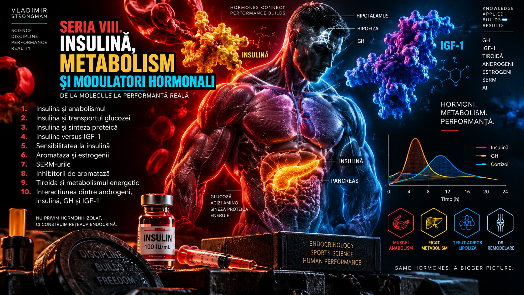
Insulin is one of the most discussed hormones in human physiology and, paradoxically, one of the most oversimplified. In nutrition conversations it is sometimes presented as “the fat-storage hormone.” In bodybuilding culture it is often called “an anabolic hormone.” In diabetology it is indispensable for survival, while in exercise physiology it is central to the way skeletal muscle handles glucose after meals and after training. Each of these statements touches one part of reality, but none of them is sufficient by itself.
To understand human metabolism, we have to abandon the idea of a single switch that decides whether energy is burned or stored. The body operates through networks. Insulin is constantly interacting with glucagon, catecholamines, cortisol, growth hormone, fatty acids, amino acids, the liver, skeletal muscle, adipose tissue and the nervous system. The final effect depends on context: fasting, feeding, exercise, recovery, sleep, body composition, insulin sensitivity and the overall metabolic state.
This first episode establishes the foundation for the new series, “Insulin, Metabolism and Hormonal Modulators.” It does not provide administration protocols and does not turn physiology into a guide for using insulin outside medical indications. Its purpose is to explain, rigorously but clearly, what the pancreas produces, how the insulin signal reaches a cell, what happens to glucose, fat and protein, and why athletic metabolism cannot be understood from blood glucose alone.
1. What is insulin, actually?
Insulin is a peptide hormone produced primarily by the beta cells of the pancreatic islets of Langerhans. Mature insulin consists of two amino-acid chains, A and B, connected by disulfide bonds. It is synthesized first as preproinsulin, processed into proinsulin, and then cleaved inside secretory granules into mature insulin and C-peptide. Insulin and C-peptide are released together, which is why C-peptide can be used clinically as a marker of endogenous insulin secretion.
Calling insulin a “glucose-lowering hormone” is correct but incomplete. Insulin lowers circulating glucose when it rises, but its biological role is much broader. It promotes the use and storage of available nutrients, inhibits several catabolic processes and coordinates the transition from the fasting state to the fed state. In practical terms, it signals that substrates are available, that some can be used immediately and that some can be stored for later.
The word “anabolic” also needs precision. In physiology, anabolic means oriented toward synthesis and building. Insulin promotes glycogen synthesis, contributes to lipogenesis in certain contexts and reduces protein breakdown. But being anabolic does not mean that every insulin rise automatically produces muscle growth, nor that every food that stimulates insulin is converted into fat. The result depends on substrate availability, energy balance and tissue responsiveness.
2. How does the beta cell decide to release insulin?
The main physiological trigger for insulin secretion is a rise in glucose. Inside the pancreatic beta cell, glucose metabolism increases the ATP-to-ADP ratio, ATP-sensitive potassium channels close, the membrane depolarizes and voltage-gated calcium channels open. Calcium enters the cell and triggers exocytosis of insulin-containing granules. It is a remarkably elegant link between nutrient metabolism, electrical activity and hormone secretion.
Glucose is not the only regulator. Amino acids, fatty acids, autonomic nervous-system activity and intestinal hormones can modify the response. GLP-1 and GIP, the incretin hormones released after nutrient ingestion, amplify insulin secretion in a glucose-dependent manner. This is one reason the same glucose load taken orally can produce a different insulin response from glucose delivered intravenously. The intestine is not merely an absorption tube; it is also an endocrine organ.
Insulin secretion also has a temporal pattern. A rapid first phase follows a sharp rise in glucose, followed by a more sustained second phase. In normal physiology, this pattern helps limit post-meal glucose excursions and coordinates hepatic and peripheral metabolism. In some forms of metabolic dysfunction, the first phase can deteriorate relatively early, before overt diabetes becomes obvious.
3. Basal versus postprandial insulin: it is not simply “on” or “off”
Insulin is present even during fasting. Basal secretion restrains uncontrolled hepatic glucose output and limits excessive lipolysis. After a meal, secretion increases and the body moves into a state of greater nutrient availability. Insulin should therefore not be imagined as a light switch with only two settings. There is a continuum of concentrations and a continuum of tissue responses.
This matters for nutrition. A mixed meal usually contains carbohydrate, protein and fat, and the metabolic response is the result of all of them together. Protein can stimulate insulin while also stimulating glucagon. Fat can slow gastric emptying and change absorption kinetics. Fiber alters the rate at which glucose appears in the circulation. There is no single “insulin response of a food” that explains the entire metabolic effect of a meal.
In an athlete, the context is even more dynamic. A meal eaten after a session that has reduced muscle glycogen reaches a muscle that is more insulin sensitive and has a genuine need to restore fuel. The same meal consumed in the setting of chronic energy surplus, inactivity and ectopic fat accumulation enters a very different metabolic system. The insulin molecule is the same; the biological terrain is not.
4. The insulin receptor: from hormone to intracellular signal
Insulin does not enter the cell to carry glucose inside. It binds to the insulin receptor on the cell membrane. The insulin receptor is a receptor tyrosine kinase. After insulin binds, the receptor changes conformation and autophosphorylates, creating docking sites for intracellular proteins such as IRS-1 and IRS-2. Several signaling pathways then branch out from this hub.
The PI3K-Akt pathway is central to many metabolic effects. Through intermediates such as TBC1D4, the signal promotes translocation of GLUT4 transporters toward the plasma membrane in skeletal muscle and adipose cells. At the same time, Akt influences glycogen synthesis, lipid metabolism, protein metabolism and transcriptional regulators. Another major branch, Ras-MAPK, has a stronger role in gene expression, growth and differentiation.
This is why insulin cannot be understood as a hormone with one effect. Its receptor starts a network. Some branches can become resistant while others remain relatively responsive, and the response differs among liver, skeletal muscle and adipose tissue. Insulin resistance is therefore not a simple condition in which “insulin stops working.” It is a complex alteration in signaling and metabolic flux.
5. GLUT4: the glucose doorway that moves to the muscle surface
GLUT4 is the glucose transporter most strongly associated with insulin action in skeletal muscle, cardiac muscle and adipose tissue. At rest, much of the GLUT4 pool is stored in intracellular vesicles. When insulin signaling is activated, these vesicles move toward and fuse with the cell membrane, increasing the capacity of the cell to take up glucose from the extracellular space.
GLUT4 transport is not an active pump pushing glucose against its gradient. It is facilitated diffusion. Insulin increases the number of available “doors,” while rapid intracellular phosphorylation and metabolism of glucose help maintain the gradient that favors entry. Once glucose enters, hexokinase converts it to glucose-6-phosphate, which can be oxidized for energy, stored as glycogen or directed toward other metabolic pathways.
For athletes, one of the most important facts is that muscle contraction can also mobilize GLUT4 through pathways that are not fully dependent on insulin. Signals involving AMPK, calcium, CaMKII and proteins such as TBC1D1 and TBC1D4 allow active muscle to increase glucose uptake even when circulating insulin is not high. Exercise does not replace insulin, but it provides an additional route for glucose disposal and can enhance insulin sensitivity afterward.
6. Skeletal muscle is a metabolic organ, not only a motor
During the postprandial period, skeletal muscle is the major site of insulin-stimulated glucose disposal. During physical activity, it becomes one of the largest glucose consumers in the body. From a metabolic perspective, muscle is not merely tissue that produces force. It is a large glycogen reservoir and a major organ of glucose homeostasis.
For a Strongman athlete, the scale of this role is amplified by muscle mass. A large amount of skeletal muscle provides substantial glycogen-storage capacity, but high muscle mass does not automatically guarantee perfect metabolic health. Chronic energy surplus, visceral or ectopic fat accumulation, inadequate sleep, certain medications, inflammation and other factors can impair insulin action even in a very strong athlete.
Training, however, provides a major metabolic advantage. Both acute exercise and chronic exercise training increase the ability of muscle to deliver, transport, use and store glucose. Modern reviews show that after exercise, insulin sensitivity can remain elevated for many hours and, in some circumstances, roughly one to two days, depending on exercise type, duration and intensity. This helps explain why regular physical activity is so powerful for glycemic control.
7. Insulin and glycogen: the fuel reservoir for repeated effort
Glycogen is the major storage form of carbohydrate in liver and muscle. Insulin promotes glycogen synthesis, but the mechanism and purpose differ between tissues. In muscle, increased GLUT4-mediated glucose transport and activation of glycogen synthase support replenishment of local stores. In the liver, glucose and insulin act together to favor storage and reduce hepatic glucose output.
In strength sports, glycogen is sometimes underestimated because a single maximal repetition is brief and depends heavily on the phosphagen system. A Strongman contest, however, is not one maximal repetition. It includes warm-ups, repeated efforts, repetition events, carries, medleys, incomplete rest periods and often many hours of physiological stress. Glycogen matters for maintaining volume and repeatedly producing high-intensity work.
Glycogen restoration is not determined solely by creating a large “insulin spike.” It depends on how much glycogen was used, how much carbohydrate is available, the time until the next session and the insulin sensitivity of the trained muscle. After exercise, the muscle is primed to replenish glycogen. The so-called post-exercise window is better understood as a period of enhanced opportunity than as a magical minute. When recovery time is short, rapid refueling matters more; when the next hard session is far away, carbohydrate distribution can be more flexible.
8. The liver: the organ that decides how much glucose enters the blood
The liver does not rely on GLUT4 as its main mechanism of glucose entry. Its metabolic logic is different. During fasting, it produces and releases glucose through glycogenolysis and gluconeogenesis, helping maintain blood glucose for the brain and other tissues. After a meal, insulin helps suppress hepatic glucose production, promotes glycogen synthesis and shifts the liver toward the fed-state metabolic program.
Hepatic control is central to understanding insulin resistance. If the liver continues to produce excessive glucose despite an adequate insulin signal, fasting glucose can rise. At the same time, other insulin-regulated pathways may remain relatively active. This selective pattern is one reason hepatic insulin resistance cannot be reduced to “insulin works” versus “insulin does not work.”
There is also important communication between adipose tissue and the liver. When insulin efficiently suppresses lipolysis, the delivery of fatty acids and glycerol to the liver declines. This reduces substrates and signaling inputs that can support gluconeogenesis and hepatic triglyceride production. Some of insulin’s effect on the liver is therefore direct, and some is mediated by changes in adipose-tissue fuel release.
9. Adipose tissue: insulin does not “create fat from nothing”
After a meal, insulin suppresses lipolysis, meaning the release of fatty acids from triglycerides stored in adipocytes. At the same time, it supports energy storage when substrates are available. This physiological fact helped generate the popular idea that insulin itself is the direct and sufficient cause of fat gain. The actual physiology is less dramatic and more important: to increase adipose mass over time, the body must store a net surplus of energy.
A person can have transient increases in insulin after meals and still lose body fat if long-term energy balance is negative. A person can also follow a diet that produces relatively modest insulin excursions and still gain fat if energy intake persistently exceeds expenditure. Insulin is a critical regulator of fuel flow, but it does not override energy conservation.
Not all fat storage is pathological either. Subcutaneous adipose tissue is a normal metabolic reservoir. Problems increase when safe storage capacity is exceeded and lipids accumulate in the liver, skeletal muscle or other organs. Ectopic lipid and lipid intermediates can interfere with insulin signaling. Healthy adipose tissue acts like a controlled fuel warehouse; when the system becomes dysfunctional, fuel begins to circulate and accumulate in the wrong places.
10. Insulin and lipolysis: a metabolic brake
Catecholamines can accelerate lipolysis in appropriate contexts, while insulin acts as a powerful brake. Through PI3K-Akt signaling and activation of phosphodiesterase 3B, insulin lowers cAMP and reduces the activation of pathways that release fatty acids from the adipocyte. During fasting and many forms of exercise, insulin concentrations fall, catecholamine influence rises and access to stored fatty acids increases.
This does not mean the body cannot oxidize fat when insulin is present. Metabolism does not use absolute gates. Synthesis and breakdown, storage and mobilization can occur simultaneously, and what matters is the net flux. After a meal, adipose lipolysis normally falls; between meals and during exercise it rises. Over the course of a day, the body continuously moves between these states.
When adipose tissue becomes insulin resistant, the anti-lipolytic effect is weakened. More free fatty acids can reach the liver and skeletal muscle, potentially worsening insulin resistance in those tissues. A metabolic feedback loop can emerge: adipose tissue responds less effectively, releases more fuel, and the excess lipid burden further disrupts insulin action elsewhere.
11. Insulin and muscle protein: “anabolic” does not mean magical
In gym culture, insulin is sometimes described as one of the most powerful anabolic hormones for muscle. Human evidence requires a more careful interpretation. A systematic review and meta-analysis of insulin studies found that one of the most consistent effects is suppression of muscle protein breakdown. Stimulation of muscle protein synthesis is more dependent on amino-acid availability and delivery.
This means insulin has an important permissive and anti-catabolic role, but essential amino acids and the mechanical stimulus from resistance training remain central to muscle protein synthesis. A lack of protein or an inadequate training stimulus cannot be compensated for simply by increasing insulin. A protein-containing meal usually produces enough physiological insulin to support the anti-catabolic role without deliberately trying to maximize insulin exposure.
For the athlete, the useful model is protein balance: muscle mass changes through the relationship between synthesis and breakdown over time. Insulin can improve that balance by reducing breakdown and, when amino acids are available, supporting synthesis. Long-term hypertrophy, however, still depends on repeated training stimuli, adequate protein and energy intake, sleep and recovery.
12. Akt, mTOR and why signaling networks matter
Akt is a central node in insulin signaling. Through several mechanisms it can influence mTORC1, a major platform regulating protein synthesis and cell growth. Yet mTORC1 does not respond only to insulin. Amino acids, cellular energy status, mechanical stress and other signals also converge on the pathway. This convergence is why no single hormone can predict hypertrophy by itself.
Activation of a pathway in a laboratory experiment is also not automatically equivalent to a meaningful long-term adaptation. A transient rise in Akt or mTOR phosphorylation after a meal or training session provides mechanistic information, but it does not guarantee greater muscle mass months later. Modern physiology tries to connect acute molecular signals with chronic outcomes, and that relationship is not always linear.
This distinction matters when “insulin hacks” are marketed. If an intervention raises insulin more for one hour, it does not follow that it will create more muscle over several months. To demonstrate that claim, actual outcomes would need to improve: muscle mass, performance and body composition, without unacceptable risk. A hormone curve is not a training result.
13. Insulin, potassium and muscle electrical function
Insulin also affects electrolytes. It stimulates Na+/K+-ATPase activity and promotes movement of potassium from the extracellular space into cells, especially skeletal muscle. This effect is strong enough that insulin, administered with glucose and other appropriate medical measures, is used in the emergency treatment of hyperkalemia. That therapeutic use illustrates how powerful the hormone can be beyond glucose regulation.
Potassium is crucial for membrane potential, muscle excitability and cardiac electrical function. An excessive fall in extracellular potassium can produce weakness and cramps and, in severe cases, arrhythmias. Insulin therefore does not merely regulate “sugar”; it also changes ion distribution.
This is another reason uncontrolled insulin use outside medical care is dangerous even before hypoglycemia is considered. A drug that can rapidly alter both glucose and potassium can affect the brain, skeletal muscle and heart within a short period. In pharmacology, the speed and magnitude of an effect matter just as much as its direction.
14. Glucagon: the partner that helps prevent blood glucose from collapsing
Insulin does not act alone. Glucagon, secreted by pancreatic alpha cells, often has opposing effects on hepatic glucose production. When blood glucose falls, glucagon supports hepatic glycogenolysis and gluconeogenesis. During fasting, the balance between insulin and glucagon helps maintain access to glucose without continuous food intake.
The insulin-to-glucagon relationship is sometimes used as a teaching shortcut to describe metabolic state. It can be useful, but real physiology also includes catecholamines, cortisol, growth hormone, substrate availability and neural signals. During hypoglycemia, epinephrine and other counterregulatory mechanisms become especially important, particularly when glucagon responses are impaired.
For athletes, this system matters during prolonged training, fasting, caloric restriction or inadequate carbohydrate intake. The fact that the body can defend circulating glucose does not mean maximal performance is guaranteed. Maintaining blood glucose for vital organs and maintaining muscle glycogen for high-level performance are different biological priorities.
15. Cortisol, catecholamines and growth hormone: counterregulatory forces
Cortisol, epinephrine, norepinephrine and growth hormone can oppose parts of insulin action, particularly during stress, fasting and exercise. Catecholamines rapidly increase fuel availability. Cortisol supports gluconeogenesis and substrate mobilization during more prolonged stress. Growth hormone has lipolytic actions and can reduce insulin sensitivity under some conditions.
This helps explain why blood glucose can rise during or after very intense exercise even while contracting muscle is consuming glucose. The liver receives strong signals to release fuel, and catecholamines rise markedly. A glucose value measured after a maximal event or a brutal training session therefore should not be interpreted as if it were a fasting morning measurement taken under standardized conditions.
Exercise physiology is a negotiation between the need to produce energy immediately and the need to restore homeostasis afterward. Insulin often falls during hard exercise, while contraction-driven pathways still permit glucose uptake. After exercise, insulin sensitivity rises and the body becomes especially efficient at restoring fuel stores.
16. What does insulin sensitivity mean?
Insulin sensitivity describes how large a biological response occurs at a given insulin concentration. If relatively little insulin can produce effective glucose uptake and appropriate suppression of hepatic glucose production, insulin sensitivity is good. If higher insulin concentrations are required to produce the same result, the system is more insulin resistant.
Insulin resistance is not one uniform disease and does not affect every organ identically. There can be predominantly muscle, hepatic or adipose insulin resistance. It can be transient in certain physiological stress states or chronic. Visceral fat, liver fat, sleep, physical activity, genetics, inflammation, medications and energy balance can all influence it.
Early in the process, the pancreas may compensate by secreting more insulin. Blood glucose may remain apparently normal while fasting or post-meal insulin is higher. This compensatory hyperinsulinemia can precede obvious dysglycemia, but individual interpretation requires context and appropriate measurements. A single fasting insulin value cannot diagnose the entire metabolic state.
17. Why does insulin resistance develop?
There is no single mechanism. In skeletal muscle, lipid intermediates, disruption of IRS-PI3K-Akt signaling, altered substrate delivery, inflammatory influences and mitochondrial dysfunction in certain contexts can contribute. In the liver, fat accumulation and lipid signaling can interfere with insulin’s control of glucose production. In adipose tissue, resistance to insulin’s anti-lipolytic action increases fatty-acid release.
Importantly, the presence of dietary fat or carbohydrate by itself does not explain insulin resistance. The relevant biology includes chronic exposure, energy flux, adipose storage capacity, physical activity, genetics and body-fat distribution. Two people of the same body weight can have very different metabolic profiles.
Assessment in very large strength athletes needs additional context. BMI becomes a poor indicator when muscle mass is extreme, but waist circumference, blood pressure, lipid profile, liver markers, fasting glucose, HbA1c, sleep quality and family history can still provide useful information. Elite performance does not exclude insulin resistance, just as one normal glucose value does not prove ideal insulin sensitivity.
18. Exercise: an insulin-independent pathway and a powerful sensitizer
Muscle contraction increases glucose uptake through mechanisms that partially converge with insulin signaling but begin from different stimuli: energetic stress, AMPK, calcium, CaMKII, mechanical signals and other molecular mediators. Active muscle can therefore consume glucose even when circulating insulin is relatively low.
The effect continues after exercise. Muscle becomes more insulin sensitive, and distal signaling elements such as TBC1D4 remain in a state that favors glucose transport and glycogen restoration. Recent reviews describe enhanced insulin-mediated glucose uptake for roughly 24 to 48 hours after some exercise protocols. Repeated training also increases proteins involved in glucose uptake and metabolism, capillary density and oxidative capacity.
For Strongman, the message is especially relevant. Heavy resistance training, carries, medleys and conditioning sessions are not only performance stimuli; they are metabolic interventions. However, enormous training stress, inadequate sleep and chronic overfeeding can work in the opposite direction. Metabolic health is the result of the entire system.
19. Blood glucose is not the same thing as insulin
Blood glucose measures the concentration of glucose in the circulation. Insulin measures the hormonal signal used to manage that glucose and other nutrients. Two people can have the same glucose concentration while requiring very different insulin concentrations to maintain it. That difference is one reason metabolism cannot be reduced to one number.
HbA1c reflects hemoglobin glycation and provides an estimate of average glucose exposure over the preceding months. It is extremely useful, but it does not directly measure insulin and can be affected by factors that alter red-blood-cell lifespan. An oral glucose tolerance test provides a more dynamic picture, and measuring insulin at the same time can add information, although interpretation is not universally standardized for every clinical situation.
In athletes, collection context matters. Recent hard training, poor sleep, infection, psychological stress, energy surplus or caloric restriction can temporarily influence markers. A serious assessment looks for patterns and trends rather than treating one value as a verdict.
20. Fasting insulin, HOMA-IR and their limitations
Fasting insulin is a simple measurement that can be useful in selected contexts. HOMA-IR combines fasting insulin and fasting glucose into an index that estimates basal insulin resistance. It has been widely used in research and epidemiology. Its main advantage is simplicity; its limitation is that it compresses complex physiology into one calculated value.
There is no perfect universal HOMA-IR cutoff. Insulin assays are not standardized identically across laboratories, and values vary with population, age, sex and method. HOMA also reflects primarily the basal relationship between hepatic glucose output, fasting glucose and insulin. It is not equivalent to directly measuring postprandial skeletal-muscle insulin sensitivity.
The hyperinsulinemic-euglycemic clamp remains a reference method in physiological research, but it is labor intensive and not a routine clinical test. In practice, simpler markers are interpreted alongside history, body composition, blood pressure, lipids and other metabolic data. An index is a tool, not a diagnosis.
21. Why “less insulin” is not always better
If insulin were judged only as a storage hormone, the logical conclusion would be that the lowest possible concentration is ideal. Biology demonstrates the opposite. Without adequate insulin, the body cannot properly control glucose production and lipolysis. In untreated type 1 diabetes, absolute insulin deficiency can lead to severe hyperglycemia, excessive ketone production, dehydration and diabetic ketoacidosis, which is life threatening.
In a healthy system, insulin should rise after eating and fall between meals. The issue is not whether a rise occurs, but whether the response is appropriate and effective. Excessive secretion required to produce a normal effect can reflect compensation. Inadequate secretion can leave glucose uncontrolled. Healthy physiology is not hormone minimization; it is context-appropriate regulation.
The same logic applies to sport. Athletes do not need to “avoid insulin” to remain lean, and they do not need to “maximize insulin” to build muscle. They need a metabolic system capable of efficiently switching between storage and mobilization, feeding and exercise.
22. Why “more insulin” is not automatically more anabolic
At the other extreme is the idea that if physiological insulin supports anabolism, more insulin must produce proportionally more muscle. This is a classic extrapolation error. Biological responses often saturate. Once key pathways are sufficiently activated and amino acids are available, raising hormone exposure further does not guarantee a proportional increase in muscle protein synthesis.
Pharmacology also introduces risks that do not exist at the same scale during normal physiology. Exogenous insulin can lower blood glucose too far and too quickly. Hypoglycemia can cause tremor, sweating, confusion, impaired coordination, seizures, loss of consciousness and death. Potassium shifts add another potential hazard.
This is the difference between explaining a mechanism and turning that mechanism into a protocol. The fact that insulin has anabolic and anti-catabolic effects does not justify use by people who do not have a medical indication. Scientific rigor requires including both the biological effect and its limits.
23. Hypoglycemia: why insulin is not a gym experiment
Current diabetes standards consider blood glucose below 70 mg/dL, or 3.9 mmol/L, clinically important and an alert threshold for hypoglycemia. Lower levels can become dangerous rapidly, especially if the person cannot recognize symptoms or treat themselves.
The brain depends heavily on circulating glucose. As glucose falls, adrenergic symptoms can appear first: tremor, palpitations, hunger and sweating. With more severe glucose deprivation, neuroglycopenic symptoms develop: difficulty concentrating, abnormal behavior, visual disturbance, confusion, seizures and loss of consciousness. Physical exercise can complicate the situation because active muscle increases glucose use.
This risk is essential in any discussion of insulin in sport. A hormone capable of causing fatal hypoglycemia should never be reduced to the casual label “nutrient partitioner.” The phrase sounds harmless; the pharmacology is not.
24. Insulin and body composition: nutrient partitioning
Nutrient partitioning describes how nutrients are distributed among oxidation, glycogen storage, adipose storage and tissue synthesis. Insulin is one component of this system, but it is not the only architect. Tissue sensitivity, training status, glycogen depletion, energy balance, muscle mass, sex hormones and genetics all influence where substrates go.
After training, a muscle that has used glycogen becomes an efficient metabolic sink for glucose. During chronic energy surplus, when glycogen is replete and intake exceeds expenditure, more energy must be stored. Even then, carbohydrate conversion to fat through de novo lipogenesis is not the only route to adipose gain. Suppression of dietary fat oxidation can also increase net fat storage.
The slogan that insulin “pushes food into muscle” is therefore incomplete. Insulin coordinates transport and storage, but the final destination of nutrients is determined by metabolic demand and the availability of all substrates.
25. Large muscle mass: a metabolic advantage, not immunity
Large skeletal-muscle mass provides a substantial reservoir for glucose and glycogen. In principle, an athlete with a great deal of active tissue has a high capacity for glucose disposal, and regular training reinforces that function. This is a real metabolic advantage.
Very large body size can also carry metabolic costs. Maintaining 130 to 160 kilograms may require enormous energy intake. If a substantial proportion of added mass is visceral or hepatic fat, if sleep is disrupted by sleep apnea, if blood pressure is high or if substances that alter metabolism are used, the muscular advantage can be eroded.
This is why metabolic health in Strongman should be treated as invisible performance. A huge deadlift does not measure hepatic insulin sensitivity. Large arms do not reveal liver fat. Performance and health can overlap, but they are not synonyms.
26. Sleep, stress and insulin sensitivity
Insufficient sleep can worsen glucose tolerance and insulin sensitivity. Mechanisms include changes in sympathetic activity, stress hormones, appetite regulation and food behavior. In an athlete, these effects may be amplified by heavy training and incomplete recovery.
Psychological and competitive stress increases catecholamines and cortisol, which can transiently alter blood glucose. A higher value after travel, four hours of sleep and competition anxiety does not carry the same meaning as a repeatedly elevated fasting value measured under standardized conditions.
This is one of the central lessons of the series: hormones are dynamic. Interpreting them without context generates myths. Metabolism is not a photograph; it is a film.
27. Insulin in sport: where physiology ends and doping begins
The insulin response after eating is normal physiology. Insulin used to treat diabetes is medicine. Administration for performance enhancement or metabolic manipulation in an athlete without a medical indication belongs to a completely different category medically and under anti-doping rules.
The 2026 WADA Prohibited List includes insulins and insulin-mimetics under S4.4.2, Hormone and Metabolic Modulators, prohibited at all times in and out of competition, subject to the relevant therapeutic-use framework when medically required. This classification is not a moral judgment about insulin. It reflects its ability to manipulate metabolism and the risks associated with misuse.
For a performance-science article, this distinction is essential. Explaining why insulin promotes muscle glucose uptake must not be converted into instructions for administration. Good science describes both the mechanism and the boundary.
28. Myth 1: “carbohydrates make you fat because they raise insulin”
Carbohydrates can raise insulin, but the claim that this rise is by itself sufficient to cause fat gain ignores energy balance. Fat loss can occur with diets containing very different proportions of carbohydrate and fat when an energy deficit is maintained. Adherence, satiety, protein intake, energy density and individual preference matter enormously.
Insulin explains why adipose lipolysis falls after a meal and why glucose is stored, but it cannot create energy. In the short term, the body can store energy after eating and mobilize it later. Over longer periods, body-mass change reflects the sum of these intervals.
For athletes, carbohydrate also serves performance. High-volume resistance work, carries, repeated events and medleys can benefit from glycogen availability. Removing carbohydrate merely to “keep insulin low” may solve an imaginary problem while creating a real one: insufficient fuel for training and recovery.
29. Myth 2: “insulin is the most anabolic hormone and should be maximized”
Insulin is anabolic in the physiological sense, but muscle hypertrophy does not increase indefinitely with insulin concentration. A meal containing adequate protein and carbohydrate normally provides enough insulin to suppress muscle protein breakdown. Amino acids and mechanical tension remain central drivers of muscle protein synthesis.
“Maximizing” a biological variable is rarely a sensible objective. Normal blood pressure is necessary for perfusion, but maximal blood pressure is not better. Insulin is necessary for metabolism, but maximal insulin is not equivalent to maximal healthy metabolism.
This basic logic is often lost in gym discussions. The body is designed around homeostasis, not hormonal records.
30. Myth 3: “if glucose is normal, metabolism is perfect”
Blood glucose can remain normal for some time even when the body secretes more insulin to keep it there. Muscle, liver and adipose insulin sensitivity can also differ from one another. One measurement cannot capture the entire system.
The opposite error is equally problematic: diagnosing “insulin resistance” from any fasting insulin result that appears high. Biological variability, laboratory method and context matter. Diagnosis belongs to medical assessment, not an online calculator.
A better approach is longitudinal: fasting glucose, HbA1c, insulin and surrogate indices when appropriate, lipid profile, blood pressure, waist measures, body weight, family history, sleep and changes over time. Metabolism is understood from a pattern, not one pixel.
31. What is metabolic flexibility?
Metabolic flexibility is the ability to change fuel use according to availability and demand. After a meal, carbohydrate oxidation can rise and adipose lipolysis can fall. During fasting and submaximal exercise, fatty-acid use can increase. During high-intensity exercise, carbohydrate becomes increasingly important because it can support high rates of ATP production.
Insulin participates in this flexibility but does not define it by itself. Mitochondria, enzymes, transporters, cardiorespiratory capacity and muscle mass all contribute. Insulin resistance is often associated with reduced metabolic flexibility, although the relationship is complex.
For Strongman, flexibility means moving from rest to explosive effort, from a ten-second event to a sixty-second medley and then back toward recovery. A high-performing metabolism is not one that burns the same fuel all the time; it is one that can change gears quickly.
32. How should metabolic testing be interpreted in a strength athlete?
A basic metabolic evaluation can include fasting glucose and HbA1c, and in selected situations a clinician may add fasting insulin, lipid profile, liver tests and other markers. If there is concern about glucose metabolism, dynamic testing or additional evaluation may be appropriate.
Standardizing test conditions helps in athletes. A maximal session, severe sleep loss, infection or an unusually large food intake can temporarily change results. Comparing values over time is more meaningful when collection conditions are similar. HbA1c may also require careful interpretation in conditions that meaningfully alter red-blood-cell turnover.
Laboratory tests should not be used as tools for self-prescribing pharmacology. Their purpose is to identify risk, follow trends and support clinical decision-making. Even an excellent biomarker becomes misleading when removed from physiology.
33. What should a Strongman remember?
First, insulin is neither the enemy created by carbohydrates nor an anabolic shortcut. It is a central regulator of the transition between fasting, feeding, exercise and recovery. Without it, glucose homeostasis fails. With efficient insulin action, muscle can rapidly use and store available fuel.
Second, training is one of the most powerful physiological ways to improve insulin sensitivity. Muscle contraction provides partially insulin-independent routes for glucose transport, while the post-exercise period favors glycogen restoration. That gives an athlete a metabolic advantage that no “insulin spike theory” can replace.
Third, size and strength do not provide metabolic immunity. A large athlete should still pay attention to waist circumference, sleep, blood pressure, lipid profile, liver health and glycemic markers. Metabolic health supports long-term performance.
34. Conclusion: insulin is a conductor, not a dictator
Insulin coordinates nutrient flow, but it does not lead the metabolic orchestra alone. It increases glucose transport in skeletal muscle and adipose tissue, promotes glycogen synthesis, suppresses hepatic glucose output, restrains lipolysis, reduces protein breakdown and alters potassium distribution. At the same time, its action is modified by glucagon, catecholamines, cortisol, growth hormone, exercise, amino acids and energy status.
Understanding insulin changes the way metabolism is viewed. There is no simple opposition between “burning” and “storage,” but a constant alternation between the two. There is no “carbohydrate is bad because it raises insulin,” but an interaction between energy balance and muscle demand. There is no “insulin is anabolic, therefore more is better,” but a dose-response relationship limited by physiology and safety.
For the strength athlete, the goal should not be to chase the lowest or highest insulin level. The goal is to build and preserve an insulin-sensitive organism that can use glucose efficiently, restore glycogen, switch between fuels and maintain metabolic health despite the extreme demands of the sport. That is the foundation on which the rest of this series will be built.
Main sources
De Meyts P. The Insulin Receptor and Its Signal Transduction Network. Endotext. A comprehensive reference for insulin receptor structure and the PI3K-Akt and MAPK signaling networks.
Weiss MA and colleagues. Insulin Biosynthesis, Secretion, Structure, and Structure-Activity Relationships. Endotext. Reference for insulin biosynthesis, proinsulin, C-peptide and beta-cell secretion.
Richter EA, Bilan PJ, Klip A. A comprehensive view of muscle glucose uptake: regulation by insulin, contractile activity, and exercise. Physiological Reviews, 2025. A modern review of GLUT4, muscle contraction and glucose uptake.
Abdulla H, Smith K, Atherton PJ, Idris I. Role of insulin in the regulation of human skeletal muscle protein synthesis and breakdown: a systematic review and meta-analysis. Diabetologia, 2016. Meta-analysis of insulin effects on muscle protein synthesis and breakdown.
Petersen MC, Shulman GI. Mechanisms of Insulin Action and Insulin Resistance. Physiological Reviews, 2018. Integrative review of insulin action in skeletal muscle, liver and adipose tissue and the mechanisms of insulin resistance.
Santos-Hernández M, Reimann F, Gribble FM. Cellular mechanisms of incretin hormone secretion. Journal of Molecular Endocrinology, 2024. Review of GLP-1, GIP and nutrient-induced intestinal endocrine signaling.
Samuel VT, Shulman GI and related literature on lipid signaling and insulin resistance. Used for the integrated relationship between ectopic lipid, hepatic metabolism and skeletal-muscle insulin action.
American Diabetes Association. Standards of Care in Diabetes 2026, sections on glycemic goals, hypoglycemia and insulin treatment. Reference for the clinical hypoglycemia alert threshold and the safety of insulin therapy.
Muniyappa R, Lee S, Chen H, Quon MJ. Current approaches for assessing insulin sensitivity and resistance in vivo. American Journal of Physiology-Endocrinology and Metabolism. Reference for the hyperinsulinemic-euglycemic clamp, HOMA and surrogate methods.
Endotext. Assessing Insulin Sensitivity and Resistance in Humans. Updated reference on the strengths and limitations of HOMA, QUICKI and direct methods.
Jaldin-Fincati JR and colleagues, together with modern reviews of GLUT4 trafficking. Used for the mechanisms of GLUT4 translocation in skeletal muscle and adipose tissue.
The 2025 Circulation Research review on skeletal-muscle insulin resistance and the restorative role of exercise. Used for the relationship between exercise training, nutrient transport and insulin sensitivity.
World Anti-Doping Agency. 2026 Prohibited List. Section S4.4.2 lists insulins and insulin-mimetics among prohibited metabolic modulators.
Editorial note: this article is educational and does not constitute medical advice or an insulin-use protocol. Insulin is an essential medicine for people with a medical indication, but inappropriate administration can cause severe hypoglycemia and other acute complications.
More From The Journal

Training
The Science of Strongman Training - Episode X: Competition Programming
How to build a complete Strongman competition preparation block: calendar, events, volume, intensity, specificity, simulations, autoregulation, tapering and fatigue management.

Training
The Science of Strongman Training - Episode IX: Peaking
Peaking in Strongman: how to turn months of training into maximal competition performance by reducing fatigue, preserving adaptations and managing specificity.