
Pharmacology
Series: The Real Cost: The Body After Pharmacology - Episode 4: Hematocrit, Viscosity and Thrombosis
September 8, 2026
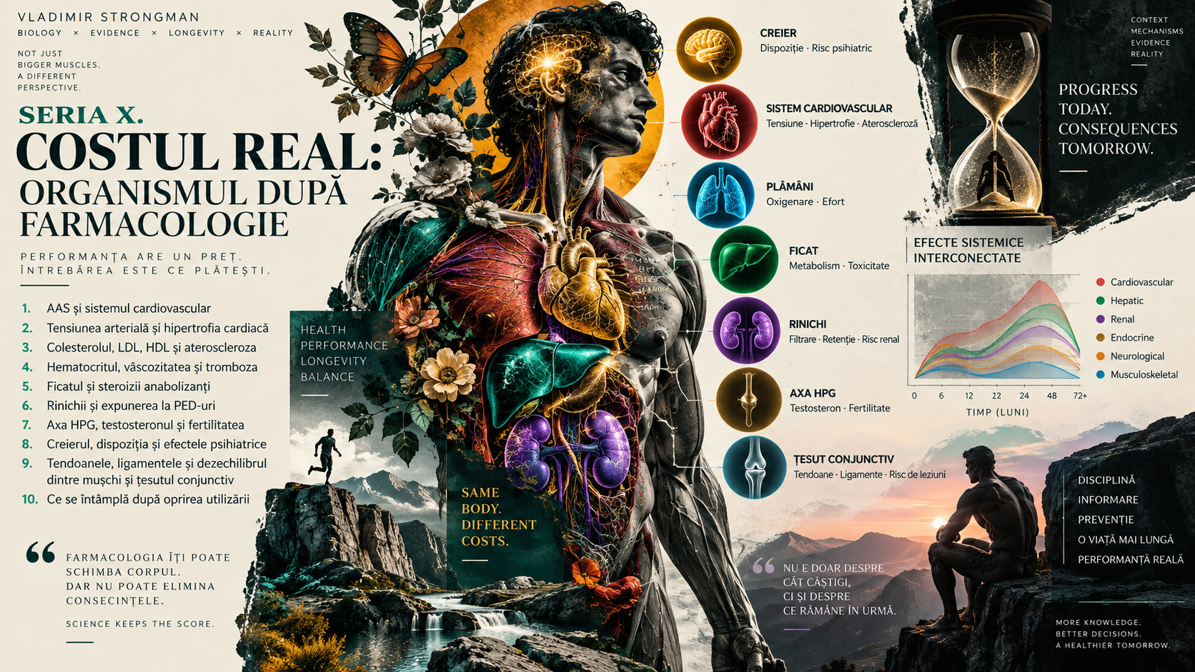
When anabolic-androgenic steroids and blood are discussed, hematocrit often becomes the main character. The reason is obvious: it appears on every complete blood count, may rise during androgen exposure and is commonly linked with the idea of “thick blood”. The reality is more complex than one number.
Hematocrit describes the proportion of blood volume occupied by red blood cells. It affects oxygen-carrying capacity, but also the mechanical properties of blood. As it rises, viscosity can increase, yet viscosity, coagulation and thrombosis are not interchangeable terms.
This episode follows the entire chain: from red-cell production and plasma volume to blood flow, endothelium, platelets, coagulation factors and clot formation. The aim is not to turn hematocrit into a villain, but to explain why risk emerges from several interacting systems.
1. What hematocrit is
Hematocrit is the percentage of total blood volume occupied by red blood cells. It can rise because red-cell mass increases or because plasma volume falls. The same laboratory value can therefore arise from very different physiological situations.
2. Hemoglobin is not the same as hematocrit
Hemoglobin is the protein inside red blood cells that carries oxygen, whereas hematocrit describes the volume occupied by those cells. The values often track together, but they are not interchangeable. Proper interpretation also considers red-cell count and red-cell indices.
3. Red blood cells are oxygen vehicles
Red blood cells carry hemoglobin and therefore most of the oxygen in blood. A moderate increase in red-cell mass can increase oxygen-carrying capacity. Beyond a certain point, however, that potential advantage can be offset by greater resistance to flow.
4. High hematocrit does not automatically mean more red cells
An elevated hematocrit may reflect increased red-cell mass, but it can also result from reduced plasma volume. This is why testing after dehydration, sauna use, heat exposure or hard exercise may look different from testing under stable conditions.
5. Plasma volume changes the picture
Plasma is the liquid component of blood. When plasma volume falls, the relative concentration of blood cells rises even without producing new red cells. In strength sports, hydration, heat and rapid changes in body mass can alter this component.
6. Hemoconcentration is different from erythrocytosis
Hemoconcentration means blood appears more concentrated because plasma has decreased. Erythrocytosis means there is a true increase in red-cell mass. Both can raise hematocrit, but the mechanism and clinical implications are not identical.
7. Absolute erythrocytosis
In absolute erythrocytosis the body truly contains more red blood cells than before. Causes can include chronic hypoxia, certain diseases, altitude exposure or androgen-driven stimulation of erythropoiesis. Diagnosis is not based on one isolated blood count.
8. AAS can stimulate erythropoiesis
Androgens can increase red-cell production through several biological pathways. The effect is well recognized even in testosterone therapy, and supraphysiological exposure may produce more pronounced responses in some individuals. The magnitude varies considerably between people.
9. Erythropoietin and hepcidin are part of the mechanism
Androgens can influence signals that favor erythropoiesis and iron availability, including pathways involving erythropoietin and hepcidin. The result can be an environment in which bone marrow produces more red cells when the required resources are available.
10. The response is not identical in every user
Some people develop substantial hematocrit increases while others show much smaller changes. Age, genetics, sleep apnea, smoking, altitude, hydration and the pattern of androgen exposure can all influence the response. A single rule applied to everyone misses this variability.
11. Altitude can raise hematocrit
At altitude, inspired oxygen pressure falls. The body adapts in part by stimulating erythropoiesis. For an athlete who combines altitude with other factors that increase red-cell mass, the whole context matters.
12. Smoking changes the equation
Carbon monoxide from cigarette smoke binds to hemoglobin and reduces effective oxygen transport. The body may compensate by increasing red-cell mass. Smoking simultaneously damages the endothelium and increases thrombotic risk through other mechanisms.
13. Sleep apnea is often overlooked
Repeated nighttime hypoxia can stimulate erythropoietic responses while increasing sympathetic activity and blood pressure. Obstructive sleep apnea is not rare in very large athletes. It can amplify several cardiovascular risk components at the same time.
14. Dehydration can raise hematocrit quickly
Water loss reduces plasma volume and can raise measured hematocrit within hours. This does not mean the marrow suddenly produced new red cells, but it can alter blood-flow properties at a time when the body is already under stress.
15. Heat adds circulatory stress
In the heat, more blood is directed toward the skin for thermoregulation and fluid is lost through sweat. If dehydration develops, plasma volume falls. For an athlete with an already elevated hematocrit, the combination can reduce cardiovascular reserve.
16. Diuretics can cause hemoconcentration
By increasing water and electrolyte loss, diuretics can reduce plasma volume and increase measured hematocrit. The concern is not just a higher number: hypotension, electrolyte disturbances, kidney injury and arrhythmias may also occur. Self-medication in this setting is dangerous.
17. Viscosity describes resistance to flow
Viscosity is a physical property of a fluid. The more blood resists deformation and flow, the harder the circulatory system must work to move it. Hematocrit is an important determinant, but it is not the only one.
18. The hematocrit-viscosity relationship is not linear
As hematocrit rises, viscosity does not necessarily increase in perfectly equal steps. At higher values, changes can become disproportionate under certain flow conditions. This helps explain why extremes are more concerning than small fluctuations.
19. Red cells must deform
Capillaries are narrow enough that red cells must deform to pass through them. Cell-membrane properties, red-cell aggregation and plasma composition affect microcirculation. “Thick blood” is therefore only shorthand for a much more sophisticated phenomenon.
20. Microcirculation is where physics becomes physiology
In small vessels, flow depends on vessel diameter, red-cell deformability, viscosity and endothelial control. A modest change in one variable may be compensated for; several unfavorable changes together can reduce tissue perfusion efficiency.
21. More oxygen carried does not automatically mean better circulation
More red cells can increase blood oxygen content, but if viscosity rises enough, flow can become more difficult. Performance depends on the balance between oxygen content and the cardiovascular system's ability to deliver it efficiently.
22. The heart sees resistance across the whole system
The heart pumps into a dynamic vascular network, not a rigid pipe. Viscosity, vascular tone and blood pressure together influence cardiovascular workload. When several rise at the same time, the hemodynamic cost can become meaningful.
23. Blood pressure and hematocrit can reinforce one another
Hypertension stresses the vascular wall and increases mechanical strain on the endothelium. More viscous blood can in turn increase circulatory resistance. Clinically, the combination matters more than either factor considered in isolation.
24. The endothelium is the active surface of blood vessels
The endothelium regulates vasodilation, inflammation, cell adhesion and coagulation. When it functions well, vessels are better able to tolerate stress. When it is dysfunctional, the environment becomes more favorable to vasoconstriction, inflammation and thrombosis.
25. Thrombosis is not simply “blood that is too thick”
A thrombus forms through activation of coagulation and interaction among platelets, endothelium and blood flow. Viscosity can influence the hemodynamic environment, but it is not the same as coagulability. This distinction is fundamental when discussing risk.
26. Virchow's triad explains the basic mechanism
The classic model of thrombosis includes three components: stasis or abnormal flow, endothelial injury or dysfunction, and hypercoagulability. Elevated hematocrit can influence parts of this model, but risk becomes more important when other factors are present too.
27. Platelets are the first response team
Platelets adhere to damaged vascular surfaces and help form the initial hemostatic plug. In pathological circumstances, the same protective function can contribute to an intravascular clot. Their activity is influenced by many factors, not just hematocrit.
28. The coagulation cascade stabilizes the clot
Coagulation factors generate thrombin and fibrin, converting the platelet plug into a more stable structure. Disturbances in this balance can favor either bleeding or thrombosis. Hematocrit does not directly measure this process.
29. Fibrinolysis is the braking system
The body not only forms clots, it also breaks them down through fibrinolysis. Thrombotic risk depends on the balance between clot formation and clot removal. A prothrombotic state can exist even when hematocrit is not dramatically elevated.
30. AAS can affect more than red blood cells
AAS exposure can affect blood pressure, lipids, endothelial function, inflammation and cardiac structure. Vascular risk should therefore not be reduced to the effect on hematocrit. Several pathways may converge in the same unfavorable direction.
31. HDL and LDL also matter in the thrombosis story
Dyslipidemia promotes atherosclerosis and endothelial dysfunction. If a vulnerable atherosclerotic plaque ruptures, platelet activation and arterial thrombosis can follow quickly. This is where episodes 3 and 4 of the series meet directly.
32. Arterial and venous thrombosis are not the same
Arterial thrombosis often develops on a background of atherosclerotic injury and platelet activation, while venous thrombosis is more strongly linked to stasis and hypercoagulability. The mechanisms overlap, but risk profiles and consequences differ.
33. Myocardial infarction can result from an arterial clot
If a thrombus blocks a coronary artery, part of the heart muscle becomes deprived of oxygen. The result can be myocardial infarction. In a young athlete, lack of previous symptoms does not exclude an underlying vascular process.
34. Ischemic stroke can have a thrombotic mechanism
A clot that obstructs cerebral circulation can cause ischemic stroke. Hypertension, smoking, atherosclerosis and other conditions can amplify the risk. Elevated hematocrit is most relevant as one element of this broader context.
35. Deep-vein thrombosis can develop in the limbs
Deep-vein thrombosis is a clot in a deep vein, commonly in the lower limb. Immobilization, surgery, inherited thrombophilia and other factors can increase risk. Intense athletic activity does not provide immunity.
36. Pulmonary embolism is an emergency
Part of a venous clot can detach and travel to the pulmonary circulation. Sudden shortness of breath, chest pain, coughing blood, dizziness or collapse requires urgent medical assessment. These symptoms should not automatically be blamed on training fatigue.
37. Immobilization after injury changes risk
An athlete may move abruptly from heavy training to days or weeks of reduced mobility after injury. Venous stasis increases in this setting. If other prothrombotic factors are present, risk may be higher than during normal training.
38. Long travel after competition is not neutral
Hours sitting in a car, bus or airplane reduce the calf-muscle pump. If dehydration, post-exercise inflammation and other risk factors are added, the context becomes less favorable for venous circulation.
39. Strongman combines several stressors in one day
A competition can combine maximal effort, Valsalva, large blood-pressure swings, heat, sweating, muscle trauma and hours of travel. Each factor may be tolerated alone; their combination is why context matters so much.
40. Dehydration should not be used to manipulate or test hematocrit
Deliberately manipulating hydration can distort laboratory values and increase cardiovascular risk. A complete blood count is more useful under reasonably stable conditions. Trends over time matter more than a value created by an artificial setting.
41. One laboratory result does not tell the whole story
Hematocrit should be interpreted alongside hemoglobin, red-cell count, hydration status and previous results. A single value can be influenced by the circumstances of the blood draw. A repeated trend is far more informative than one isolated result.
42. The complete blood count is the starting point
A complete blood count provides hematocrit, hemoglobin, red-cell count and red-cell indices. It can suggest erythrocytosis, iron deficiency or other hematologic problems. The numbers need to be interpreted with the clinical picture rather than treated as a scoreboard.
43. Iron and ferritin can change interpretation
Increased red-cell production consumes iron. If stores fall, red-cell indices can change and iron-deficiency symptoms may develop. Repeated manipulation of blood volume without supervision can make the situation more complicated.
44. Erythropoietin can help investigate the cause
In selected situations, clinicians may use erythropoietin levels and other tests to distinguish causes of persistent erythrocytosis. The goal is to determine whether the problem is secondary to hypoxia or hormonal exposure, or whether a hematologic disorder needs to be considered.
45. Persistent elevation requires consideration of hematologic disease
Not every high hematocrit in an AAS user should automatically be blamed on AAS. Polycythemia vera and other disorders can cause erythrocytosis and have specific implications. An apparently obvious explanation should not stop investigation when abnormalities persist.
46. Oxygen saturation can provide clues
Low oxygen saturation or nocturnal desaturation can suggest hypoxia as a stimulus for erythropoiesis. Pulse oximetry has limitations and does not replace clinical evaluation, but it can be one useful piece of the puzzle.
47. Sleep apnea deserves investigation when suspected
Loud snoring, witnessed pauses in breathing, daytime sleepiness and unrefreshing sleep can suggest obstructive sleep apnea. Treating nocturnal hypoxia can have effects beyond hematocrit, including benefits for blood pressure and overall cardiovascular risk.
48. Routine coagulation tests do not measure all thrombotic risk
PT, INR and APTT are useful in specific settings, but normal results do not prove the absence of thrombosis risk. Thrombosis is influenced by factors that standard coagulation tests do not capture completely.
49. D-dimer is not a screening test for every athlete
D-dimer can be useful in clinical algorithms when thromboembolism is suspected, but it can rise in many other circumstances. Used without context, it may create confusion. Testing decisions should be based on symptoms and clinical probability.
50. Some values require medical review rather than improvisation
In testosterone-therapy guidelines, hematocrit around or above 54% is treated as an important signal requiring reassessment of treatment and associated causes. That threshold should not be turned into a universal rule for illicit AAS use or a self-treatment protocol.
51. Blood donation is not a universal solution
Removing blood can temporarily lower hematocrit, but it does not correct the cause driving it and repeated removal can deplete iron stores. Therapeutic phlebotomy is a medical intervention whose indication depends on diagnosis, not on forum advice or a single number.
52. Aspirin should not be used automatically
The fact that aspirin reduces platelet aggregation in certain cardiovascular diseases does not make it appropriate for every person with elevated hematocrit. It can cause bleeding and has specific medical indications. Self-medication can simply trade one risk for another.
53. Hydration corrects hemoconcentration, not true erythrocytosis
If hematocrit is high because plasma volume has fallen, restoring hydration may change the measurement. If red-cell mass is genuinely increased, drinking more water does not remove the cause. Confusing the two situations produces many false conclusions.
54. Improving one factor does not erase the others
Even if hematocrit returns to a better range, hypertension, dyslipidemia, smoking, sleep apnea or atherosclerosis may remain. Cardiovascular risk management does not work by fixing a single number.
55. Symptoms may be absent
Elevated hematocrit can exist without obvious symptoms. Headache, dizziness or a sense of pressure are neither sensitive nor specific. Feeling normal cannot be used as proof that risk is absent.
56. Some symptoms are red flags
Chest pain, sudden shortness of breath, one-sided weakness, speech disturbance, fainting or painful unilateral leg swelling require urgent medical assessment. This is not the setting for experiments with hydration, supplements or unsupervised medication.
57. Trends matter more than hunting for one threshold
A progressive rise in hematocrit accompanied by increasing blood pressure, worsening lipids or symptoms is more informative than mechanically comparing one result with a laboratory cutoff. Longitudinal monitoring shows the direction in which the system is moving.
58. Stopping exposure may allow reversibility
If androgen stimulation is the main driver, hematocrit can gradually decline after exposure is reduced or stopped as red cells age and are removed from circulation. The change is not immediate, and other causes may keep the value elevated.
59. Risk is an equation, not a single number
Hematocrit, viscosity, blood pressure, lipids, smoking, sleep, hydration, immobilization and individual predisposition can combine. No single parameter should be treated as sacred. Cardiovascular medicine deals with probabilities and contexts, not numerical talismans.
60. Conclusion: “thick blood” is only the beginning of the explanation
Elevated hematocrit can increase viscosity and add strain to an already stressed cardiovascular system, but thrombosis is not created by one laboratory value. It emerges when flow, endothelium and coagulation meet in an unfavorable context. That is why assessment must be integrated.
Viewed simplistically, hematocrit looks like a measure of how much “red blood” a person has. Viewed correctly, it sits at the intersection of red-cell mass and plasma volume. That distinction explains why dehydration and true erythrocytosis should not be confused.
Viscosity is a relevant physical consequence, but it does not automatically turn blood into a clot. Thrombosis requires a much more complex biology involving platelets, coagulation, vulnerable endothelium and altered flow.
AAS can contribute to this equation through several pathways at once. They may increase erythropoiesis in some people while also affecting blood pressure, lipids, endothelial function and cardiovascular structure. The accumulation of effects matters more than any one pathway in isolation.
For the strength athlete, competition context matters enormously. Heat, fluid loss, Valsalva, post-exercise inflammation, injury and long travel can turn a tolerable resting situation into one with far less physiological reserve.
The central message of this episode is that useful monitoring does not search for a magic number. It follows trends, causes and interactions. Elevated hematocrit should be understood, not cosmetically corrected with improvised solutions.
Sources and recommended reading
1. Guyton and Hall, Textbook of Medical Physiology: red-cell physiology, hematocrit, circulation and blood viscosity.
2. Boron & Boulpaep, Medical Physiology: oxygen transport, microcirculation and the rheological properties of blood.
3. Endocrine Society Clinical Practice Guideline on testosterone therapy in men with hypogonadism: hematocrit monitoring and management of erythrocytosis in therapeutic use.
4. American Urological Association, Testosterone Deficiency Guideline: recommendations on hemoglobin, hematocrit and monitoring during testosterone therapy.
5. Hematology literature on secondary erythrocytosis and polycythemia vera: differential diagnosis, erythropoietin and evaluation of persistently elevated hematocrit.
6. Virchow and modern literature on Virchow's triad: stasis, endothelial injury and hypercoagulability as a framework for thrombosis.
7. European Society of Cardiology and contemporary guidance on venous thromboembolism and pulmonary embolism: mechanisms, diagnosis and risk assessment.
8. Physiology and hemorheology studies on hematocrit, viscosity, red-cell deformability and microcirculation.
9. Clinical literature on AAS and the cardiovascular system: effects on erythropoiesis, blood pressure, endothelium, lipids and vascular risk.
10. Sports-medicine reviews on dehydration, heat exposure, travel, immobilization and thrombotic considerations relevant to athletes.
Editorial note: this material is educational and does not provide protocols for AAS use, phlebotomy, antiplatelet drugs or anticoagulants. Persistently elevated hematocrit, symptoms suggesting thrombosis or cardiovascular abnormalities require individualized medical assessment.
More From The Journal

Training
The Science of Strongman Training - Episode X: Competition Programming
How to build a complete Strongman competition preparation block: calendar, events, volume, intensity, specificity, simulations, autoregulation, tapering and fatigue management.

Training
The Science of Strongman Training - Episode IX: Peaking
Peaking in Strongman: how to turn months of training into maximal competition performance by reducing fatigue, preserving adaptations and managing specificity.