
Sports Medicine
Series: STRONGMAN INJURIES: FROM TRAUMA TO RETURNING TO COMPETITION - Episode 6: Muscle Injuries and the Biology of Healing
October 12, 2026

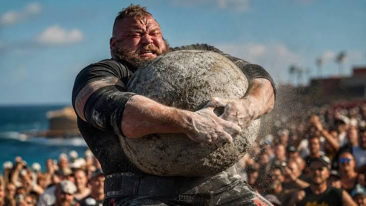
In a strongman competition, a muscle injury does not always arrive with the dramatic image of a complete tear. An athlete picks up a yoke, then feels a sharp pain in the thigh on the third step. Another tries to accelerate with farmers implements and suddenly cannot push effectively from one leg. During a loading medley, a stone must be brought into the lap and a competitor experiences sudden groin pain. Sometimes these symptoms reflect structural damage; at other times they involve a functional response to load without an identifiable macroscopic fibre tear. For an athlete whose sporting identity depends on producing extraordinary force, both situations may mean weeks of disrupted preparation. Distinguishing between them is essential to diagnosis, treatment, and a responsible return to competition.
The sixth episode of “Strongman Injuries: From Trauma to Returning to Competition” explores muscle injuries and the particular biology of healing. We will not portray muscle as a rope that snaps and is subsequently glued together, but as living tissue in which contractile fibres, connective structures, blood vessels, immune cells, and the nervous system participate in dynamic regeneration. We will distinguish delayed-onset muscle soreness from strains, contusions, partial tears, and complete ruptures, then follow rehabilitation from the first hours through the return to maximal events. Deadlifts, yoke carries, farmers walks, atlas stones, sandbags, log presses, and tyre flips provide relevant sporting examples, without inventing event-specific injury rates that research has not established.
The everyday phrase “muscle tear” can conceal injuries that differ substantially in anatomy, severity, and prognosis. Some primarily affect muscle fibres; others involve the transition between muscle and tendon, and the more complex injuries may extend into tendinous structures located inside the muscle. The same painful region may therefore involve different rehabilitation requirements. Not every severe pain during exertion indicates a rupture, and not every abnormality visible on MRI requires surgery. The Munich consensus attempted to address this ambiguity by distinguishing functional muscle disorders without identifiable macroscopic tears from structural injuries involving muscle fibres [2]. That terminology supports communication, but it is an expert consensus rather than proof that an imaging label can predict the exact day of recovery.
Why are muscles particularly important in strongman? Because they do not merely create force in a single direction. They accelerate and decelerate loads, maintain energetically costly positions, control unstable implements, and repeat near-maximal contractions under fatigue. In the same session, the posterior thigh muscles may be challenged by deadlifts, the quadriceps by yoke carries and squats, the adductors during stone loading, and the shoulder-girdle muscles during heavy log pressing. This variety creates a need for thoughtful programming, but it does not mean every fatigued muscle is about to tear. Sports medicine must distinguish plausible biological mechanisms from injury risks quantified by research.
Skeletal muscle: the structure behind strength
Skeletal muscle contains numerous muscle fibres, each housing myofibrils and contractile units known as sarcomeres. Inside these units, actin and myosin filaments interact to generate tension. Fibres are organised into fascicles and surrounded by connective tissue, while blood vessels and nerves supply oxygen, metabolic substrates, and motor control. This architecture explains why an injury is more than the problem of a single torn fibre. Connective structures, local circulation, and the way force travels through muscle may be affected simultaneously.
The endomysium surrounds individual fibres, the perimysium organises fascicles, and the epimysium contributes to the muscle's outer covering. These structures are not merely packaging: they participate in mechanical organisation and force transmission. In certain injuries, involvement of connective tissue or intramuscular tendons has different significance from a primarily peripheral muscular lesion. The British Athletics Muscle Injury Classification, introduced by Pollock and colleagues, uses MRI-based grades and suffixes to describe myofascial, musculotendinous, or intratendinous locations [3]. It is a method of description and stratification, not an infallible countdown to competition.
The musculotendinous junction is the interface where contractile components meet tendinous structures and where particular combinations of force can produce local stress. In many sprinting and rapidly accelerating sports, injuries occur near this interface, although their distribution cannot automatically be transferred to strongman. A heavy deadlift and a maximal sprint challenge the hamstrings in different ways; a stone lifted from deep hip flexion involves yet another combination of positions and contractions. The general anatomical lesson is that the integrity of the whole muscle–tendon unit and its ability to transmit force matter more than visible muscle size alone.
Muscles do not develop tension only while shortening. During concentric action a muscle generates force as it shortens; during eccentric action it resists force while lengthening; during isometric action overall length changes little while tension is maintained. All three types occur in strongman. Lowering an object under control can create eccentric loading, holding implements involves considerable isometric effort, and lifting relies on concentric work. Eccentric contractions are valuable for strength development and can form part of rehabilitation, but unfamiliar exposure may provoke delayed-onset soreness. That response does not automatically mean traumatic fibre rupture.
Soreness, overload, and rupture: three different things
Delayed-onset muscle soreness, or DOMS, usually follows unfamiliar exercise or substantial eccentric loading. It is neither an obligatory sign of progress nor proof that the athlete has sustained a major structural injury. It often involves diffuse tenderness, stiffness, and a temporary reduction in performance, beginning hours after exercise and commonly becoming more noticeable over the following one or two days. A competitor who suddenly changes exercises, greatly increases repetitions, or introduces a previously unfamiliar event may experience DOMS without requiring treatment for a torn muscle. Evolution and context matter more than the gym expression “I completely destroyed my legs.”
A structural muscle injury may begin suddenly during effort and be accompanied by focal pain, a tearing sensation, reduced strength, and sometimes bruising or a palpable defect. These findings raise suspicion without establishing the injury grade. During the first hours, pain and swelling can limit examination, while bruising may develop later and appear away from the original site as blood tracks through tissue planes. A person may still walk despite a substantial tear, just as a relatively small lesion may be intensely painful. Diagnosis depends on the specific history, functional examination, and appropriate investigations when they may change management.
In Munich-consensus terminology, functional muscle disorders can cause symptoms without a macroscopic fibre rupture visible through available imaging [2]. They are neither imaginary nor necessarily unimportant. An athlete may genuinely struggle with acceleration, hip extension, or maintaining posture even when MRI reveals no fibre discontinuity. Nonetheless, the classification is broad and largely represents an expert framework. Clinicians must consider alternative explanations, including referred pain, neural involvement, tendon disorders, and systemic disease. The absence of a visible rupture does not automatically end the diagnostic process.
A contusion has a different mechanism: a direct impact damages local tissues and may produce bleeding. A dropped implement striking the thigh, a collision with a yoke frame, or a heavy object pressing forcefully against the arm can create such injuries. Severe contusions can involve large haematomas and loss of function, with rare circumstances requiring investigation for complications such as compartment syndrome. Aggressive deep massage immediately after substantial trauma and swelling should not be presented as routine treatment. Assessing severity and initially protecting the region may be more important than trying to accelerate a return to training.
Complete ruptures and avulsions require separate consideration. When a muscle or its attachment loses continuity to a major extent, the resulting functional deficit can be substantial. Selected proximal hamstring avulsions, markedly retracted tears, and other specific injuries may warrant prompt surgical assessment. Not every complete rupture requires surgery, and not all partial tears have the same prognosis. Location, degree of retraction, athletic requirements, and time since injury are important factors. In strongman, confusing “I can still bend my leg” with “I can safely produce maximal force again” is a recipe for premature decisions.
Injury classification: what a grade tells us and what it cannot
The familiar grade I, II, and III system provides a rough description of severity but can be too crude when it ignores location and the structures involved. Two injuries both described as grade II may differ dramatically in extent, position, and sporting demands. The Munich consensus distinguishes functional disorders from partial and complete tears [2], while the British Athletics approach adds MRI grading and location relative to the fascia and intramuscular tendon [3]. Neither system removes the need for examination, and neither has been validated as a universal tool for predicting exact time loss in every sport.
MRI can describe features visible at a particular moment: oedema, signal changes, fibre discontinuity, haematoma, or tendon involvement. Ultrasound can provide dynamic information and may be useful in some superficial injuries, but findings depend on equipment, operator expertise, and timing. Very early in an injury, the absence of conspicuous imaging changes does not always exclude a clinically important problem. Conversely, a residual signal abnormality after symptoms improve does not automatically prove that an athlete cannot return to activity. Imaging results contribute to diagnosis; they do not replace functional assessment.
Classification is especially valuable when it improves communication. Instead of saying “he pulled his hamstring,” a team can discuss whether the lesion is proximal or distal, whether a tendon is involved, which contractions reproduce pain, and which movements remain limited. Those details may influence exercise selection and the pace of reloading. Nevertheless, an elegant diagnostic label does not substitute for actual progress. Some individuals recover faster than images might suggest, while others retain weakness or fear after initial pain has settled. Rehabilitation should be updated according to the athlete's response rather than reciting a classification mechanically.
How muscle heals: inflammation, regeneration, and remodelling
Muscle repair begins immediately after injury. The affected area may undergo fibre disruption, bleeding, alterations of the extracellular matrix, and recruitment of immune cells. For teaching purposes, research often divides healing into an initial destruction and inflammatory phase, a regenerative phase, and a remodelling phase [4,5]. In the body, these processes do not follow one another as three perfectly separate chapters: they overlap and vary with injury severity, location, and the individual's biological condition. This overlap explains why there is no calendar on which every athlete crosses from “inflammation” into “fully healed muscle” on precisely the same day.
Satellite cells, located close to muscle fibres, are muscle stem cells that contribute to repair and regeneration. Following injury, biological signals can activate them, leading to proliferation and participation in restoring contractile structures. The process also involves immune cells, blood vessels, fibroblasts, and remodelling of the extracellular matrix. The statement “muscle grows back by itself” captures only part of this biology. Functional recovery requires sufficiently effective organisation of contractile fibres and supporting connective tissue to tolerate tension and transmit force again. Regenerative capacity is remarkable, but it is not limitless [4,5].
Early inflammation helps remove damaged material and signals later stages of tissue repair. This does not mean every episode of swelling should go unmanaged, or that all anti-inflammatory medication must be prohibited. It means inflammation is not exclusively an enemy and that interventions should be assessed against symptoms, benefits, and risks. Analgesia may have a role in individual care, but eliminating pain temporarily is not a test of healing. Nor is there good evidence that one standard package of massages, creams, and devices can predictably accelerate every biological stage in every muscle injury.
Following more substantial injury, connective-tissue repair can produce fibrosis. Scar tissue is not automatically a sign of failure, because connective tissue helps stabilise the injured region. Excessive fibrosis or poorly organised repair can, however, affect mechanical properties and tolerance of future loading [5,15]. This possibility should not become a universal explanation for pain at the site of an old injury. An ultrasound appearance consistent with scarring does not by itself reveal how much force an athlete can generate. Clinical relevance emerges when structure, function, and symptoms are considered together.
Remodelling continues after acute pain subsides. An athlete may walk normally or perform light contractions before tissue is ready for explosive repetitions, directional changes, or maximal efforts. Rehabilitation therefore requires more than waiting a prescribed number of days. It introduces relevant tension progressively at an appropriate pace. Insufficient activity can delay reconditioning, while abrupt exposure can exceed current capacity. The goal is not to choose between complete rest and maximal strength, but to construct a practical bridge between them according to the condition and the sport's demands.
Vulnerable regions in strongman: why the event changes the mechanism
The hamstrings, located at the back of the thigh, contribute to hip extension and, with anatomical differences between individual muscles, knee flexion. Deadlifts and many hip-hinge variations challenge them at different muscle lengths. A heavy pull from the floor is not biomechanically identical to maximal sprinting, although both can create considerable hamstring demand. Most hamstring injury research comes from running-based sports; it provides valuable principles of muscle rehabilitation but does not demonstrate the same distribution of injuries in strongman [6,7]. A competitor with posterior thigh pain should be evaluated according to the task that provoked it, rather than diagnosed purely from football injury statistics.
The adductor muscles help position and stabilise the legs under load. During atlas stone and sandbag loading, a wide stance and rapid transitions between hip flexion and extension can change local muscle demands. During yoke carries, each step also requires frontal-plane pelvic control rather than knee extension alone. Groin pain can arise from muscles, tendons, joints, or non-musculoskeletal conditions. Calling it an “adductor tear” without examination can delay the correct diagnosis. Especially after a sudden episode associated with loss of strength, the athlete must distinguish ordinary discomfort from a problem that justifies stopping the event.
The quadriceps faces considerable demands during squats, yokes, sled pushes, truck pulls, and stone-loading phases. It can be affected by direct injury or indirect loading, but muscle injuries must be distinguished from quadriceps and patellar tendon ruptures, discussed in the previous episode. If a competitor develops sudden severe pain at the front of the thigh, the clinically useful questions concern active knee extension, palpable defects, recent impact, bruising, and loss of force. These observations cannot provide a complete diagnosis, but they help determine urgency. A severe contusion with internal bleeding is not treated like ordinary soreness after a novel squat session.
The gastrocnemius and soleus contribute to ankle control and the push against the ground, including during farmers runs and vehicle pulling. Athletes sometimes underestimate the calf muscles because they are not directly associated with maximal deadlift records, but every fast step under load requires force and impulse control. Sudden calf pain can indicate muscular injury, but vascular problems occasionally need to be excluded, especially when there is marked unilateral swelling or relevant risk factors. It is unwise to recommend aggressive stretching or deep massage for an acutely painful calf before the mechanism and associated symptoms have been properly assessed.
Back muscles also play a central role, yet lower-back pain must not automatically be called an “erector spinae tear.” Paraspinal muscles can become sensitive after exertion, show protective muscular responses, or less commonly sustain structural injuries. The source of pain may instead involve discs, joints, nerves, or other structures, as discussed in episode four. Coherent clinical assessment considers location, neurological signs, movement response, and symptom evolution. Numbness, progressive weakness, or new bowel or bladder problems make an improvised diagnosis of “muscle spasm” particularly dangerous. Strongman needs medical literacy, not absolute certainty based on a single sensation.
In overhead events, the deltoids, pectorals, triceps, and scapular muscles may face sharp peaks of tension, especially when an implement deviates and the athlete tries to regain control. Sudden chest pain accompanied by deformity and bruising can raise suspicion of a pectoralis injury, but assessment must determine whether the injury involves muscle, the musculotendinous region, or tendon. Treatment and prognosis differ. Likewise, arm pain after handling a stone or tyre does not automatically confirm a biceps rupture. The third episode explained why proximal and distal tendon injury locations change management. The same anatomical precision is needed for muscular injuries.
Strongman injury risk cannot be reduced to a map of “weak muscles.” Performance often requires adaptation to unusual implement dimensions and centres of mass, transitions made against the clock, and steps taken under fatigue. These conditions can alter the rate of force development and coordination, but research cannot provide robust estimates for each muscular injury during each event. We should therefore distinguish physiologically plausible mechanisms from demonstrated causes. An athlete injured during a sandbag carry cannot establish that it is more dangerous than deadlifting merely because one injury occurred in that sequence.
Medical assessment and imaging: when investigations are useful
Assessment begins with a history: was there a distinct incident, impact, acceleration, change of position, or gradual deterioration in tolerance? Where is the pain, when did it begin, does it occur at rest or only during contraction, and are swelling, bruising, or visible changes in muscle contour present? Previous injury, recent training volume, unfamiliar exercise, accumulated fatigue, and medication are also relevant. Clinical examination includes inspection, appropriate palpation, mobility testing, and strength assessment within safe limits. After a significant acute injury, attempting a maximal deadlift or squat is not a legitimate diagnostic test.
Ultrasound can be useful for selected muscle injuries, haematomas, fibre discontinuity, and local follow-up. MRI provides a broader view of deep injuries, intramuscular tendon involvement, and alternative diagnoses, but it is not automatically required for every painful episode. The investigation should answer a clinical question: is there a tear large enough to change treatment, an avulsion that may need surgery, a significant haematoma, or another condition? An image confirming a small area of oedema without meaningful functional consequences may contribute little to an otherwise well-founded rehabilitation plan.
An imaging classification must not be confused with the athlete's actual physical capacity. Hamstring guidance emphasises a combination of clinical examination, symptoms, restored strength, and sporting requirements [6,7]. Predicting the exact return date and reinjury risk from MRI appearance alone remains difficult. This limitation is even greater for strongman, where an athlete may be ready for everyday movement but not for controlling a maximal yoke or an awkward stone. Data from football concerning return to sprinting do not establish deadlift weights or farmers-walk distances. They provide a starting point for clinical reasoning, not a direct prescription.
Sometimes investigation is urgent. A suspected complete rupture, rapidly expanding haematoma, major loss of function, unusually severe pain after trauma, or neurovascular abnormalities warrant prompt assessment. Pain accompanied by fever and systemic illness must be considered differently from an ordinary strain. With severe pain, an increasingly tense muscular compartment, and worsening symptoms, acute compartment syndrome must be considered. This is an emergency that cannot be safely excluded through a quick impression in the gym [13]. In these circumstances, medical care takes priority over completing a training programme.
The first 24–72 hours: protection without unnecessary immobilisation
Immediately after a suspected structural muscle injury, the athlete should stop the provoking exertion and have the severity assessed. Temporary protection can reduce the chance of aggravating an injury, but it does not automatically imply prolonged total rest. In many uncomplicated presentations, retaining tolerable activity and gradually reintroducing movement are useful aspects of recovery. Familiar acronyms such as RICE, PRICE, and PEACE & LOVE attempt to summarise care principles, but they are not uniformly validated protocols for every muscle tear. The PEACE & LOVE editorial emphasises education and gradual loading; it should be treated as conceptual guidance rather than a clinical trial proving an approach for every strongman trauma [16].
Ice may temporarily reduce pain perception in some settings, but there is no strong evidence that its application accelerates all stages of muscle regeneration in every case. Compression and limb positioning may help manage selected symptoms, provided they do not obscure deterioration or become excessive. Major trauma, enlarging haematoma, or disproportionate pain requires an individual medical decision. Deep massage in the first hours following a painful bleeding injury is not a mandatory intervention and may be inappropriate. The early objective is to protect function and establish what has happened, not to rush into local tissue manipulation.
Painkillers and anti-inflammatory medications require separate consideration. Some can reduce symptoms, but their risk profiles differ and may include gastrointestinal, kidney, and cardiovascular complications. The relationship between inflammation and tissue regeneration is biologically complex, and that complexity does not prove that every anti-inflammatory medication should be universally banned. Decisions must account for severity, general health, other medications, and professional medical advice. Taking substances to conceal pain before another heavy event creates a different problem: useful symptoms may be suppressed without repairing injured tissue. Masked pain is not proof of healing.
During the first days, athletes may notice that one movement is tolerable while another is not. This can help with temporary programme adjustments, but it must be interpreted carefully. After an injury that may require intervention, pain decreasing over several hours does not guarantee the problem has resolved. After a minor injury has been assessed appropriately, walking and other comfortable activities may help preserve function. The distinction is based on diagnosis and functional response, not determination. Being able to use a limb in ordinary life does not demonstrate readiness for hundreds of kilograms.
Conservative care and indications for surgery
Many muscle injuries can be treated without surgery through appropriate protection, load adjustment, and progressive rehabilitation. Conservative care does not mean doing nothing. Physiotherapy may aim to restore range of motion, strength, coordination, and tolerance of contractions at different muscle lengths. Large ruptures, avulsions, substantial retraction, or marked functional deficits may require specialist assessment to determine whether surgery offers a meaningful advantage. There is no universal rule that every tear needs an operation, nor can anyone promise that every rupture will heal fully through exercise alone.
In a proximal musculotendinous avulsion, for example, the precise location and extent of injury can change treatment goals. Some patients recover through rehabilitation, while others may be candidates for surgical repair; postoperative recovery then involves specific restrictions. Recommendations for a minor muscle strain should not be applied directly to a complete avulsion. For strongman, a loss of maximal force may matter greatly even when the person can walk and climb stairs normally. Clinical discussion should therefore include competitive demands rather than merely the minimum level of function needed for daily life.
Another mistake is assuming surgery always offers the shortest route back. Every operation has risks, healing requirements, and a rehabilitation process. In addition, not every persistent symptom comes from a structure that can be fixed mechanically. Accurate diagnosis and a discussion of treatment options are essential. The best choice for an athlete is not necessarily the one that permits the quickest light training session, but the one offering a reasonable long-term functional outcome. Decisions belong to the individual and medical team, informed by evidence and clinical details.
Functional rehabilitation: progressive exposure to tension
Rehabilitation may begin with gentle movement and low-load contractions, then progress through adapted strengthening, eccentric demands, and eventually sport-specific conditions. This sequence is not a fixed recipe; injury type, pain, structural integrity, and clinical advice matter. Early goals often include preserving appropriate motion and avoiding needless loss of conditioning. Later stages require strength at relevant muscle lengths, repeatable force production, and the ability to generate or absorb force at the speeds needed in sport. In strongman, being pain-free during a gentle exercise is not equivalent to readiness for a complete loading medley.
Isometric exercise may allow muscular activation without major joint movement and can be well tolerated in certain cases. Concentric and eccentric work introduce different demands, while loading at longer muscle lengths may become relevant after capacity returns. Studies of hamstring injuries have examined lengthening exercises and progressively demanding rehabilitation, primarily among football players [8,9]. These findings do not mean every strongman must start aggressive eccentric work on the first day. They suggest that rehabilitation must restore actual function and cannot be reduced to massage and inactivity.
One randomised trial compared earlier with delayed introduction of lengthening exercises in male athletes with acute hamstring injuries. The difference in return-to-sport time was not statistically significant, and earlier introduction did not demonstrate a clear advantage regarding reinjury [9]. This is useful because it challenges commercial messages suggesting that “earlier is always better.” Progressive loading is important, but the timing and selection of exercises must be individualised. Sometimes the most productive decision is to consolidate one stage before introducing another challenge.
The London international consensus on hamstring rehabilitation recommends programmes tailored to the athlete, the muscles involved, injury type and severity, and specific sporting demands [7]. Progression criteria, symptom response, and gradual exposure to competition-relevant work matter. Experts did not agree on every strength and flexibility benchmark, and the consensus does not provide a universal numerical threshold for every case. In strongman, uncertainty is even greater: criteria developed for returning to sprinting cannot simply be converted into deadlift kilograms, yoke distances, or the number of stones to load.
A useful practical principle is not to increase every aspect of loading simultaneously. When reintroducing Romanian deadlifts following hamstring injury, it can help to consider range of motion, load, repetitions, and movement speed separately. For an injured adductor, stance width, hip range, and pelvic control may matter. After a quadriceps contusion, knee-flexion tolerance and extensor strength may be relevant. These are programming principles rather than universal medical protocols. Their application depends on the diagnosis and the athlete's response during and after training.
Pain is not the only outcome. Rehabilitation may assess range of motion, focal tenderness, contraction strength, ability to repeat exertion, walking or running where relevant, and responses over the next 24–48 hours. In selected musculoskeletal conditions, limited transient discomfort may be compatible with supervised exercise; in other situations, painful loading is inappropriate. No pain score authorises all exercises following every muscle tear. Newly developing weakness, a growing haematoma, or neurological symptoms changes priorities and warrants reassessment.
Returning to deadlifts, yokes, farmers walks, stones, and log presses
Returning to deadlifts after a hamstring or other hip-extensor muscle injury should not be defined solely by the ability to lift an empty bar from the ground. Over time, the athlete needs to demonstrate tolerance of relevant range of motion, increasingly forceful contractions, repeated effort, and recovery between sessions. A limited-range hip-hinge exercise may precede a conventional deadlift, followed by gradual loading without turning every session into a maximal test. If an avulsion has surgical indications or specific restrictions, progression must be adapted under professional supervision. This example describes principles, not universal percentages of a previous personal best.
During yoke carries, the lower-limb muscles must tolerate the pick-up and repeated stepping, acceleration, and corrections to frame oscillation. An athlete recovering from a quadriceps or adductor injury may manage a static hold before tolerating a fast carry. Progression can separate the initial pick-up, holding the frame, taking controlled steps, and gradually increasing distance or speed. Even after pain disappears, sharp directional changes and unexpected disruptions may remain unfamiliar demands. Complete return should reflect the actual event, not merely a perfect repetition performed in controlled conditions.
Farmers walks and frame carries repeatedly load the calf, thigh, hip, and trunk muscles. After injury, an athlete may be tempted to reduce only the weight while retaining the usual long distance and fast pace. Yet movement speed, step rhythm, or total exposure time may be the most relevant symptom triggers. A clearer strategy may modify one variable while observing the response. Reintroducing turns, picking up the implements, and lowering them safely matters just as much as the central portion of a run. No isolated test can guarantee that an injury will not recur.
Atlas stones and sandbags require deep hip positions, bringing awkward objects close to the body, and transitions between phases. These demands affect adductors, glutes, and the posterior chain differently from conventional lifts. Someone who can perform a pain-free hip thrust may still be unprepared to lift a bulky stone from the floor. Reintroduction may begin with lighter, more predictable implements, then add event-specific transitions, further repetitions, and finally conditions resembling competition. Loading choices should reflect the diagnosis and the response between sessions, not the assumption that any stone below half the personal best is automatically safe.
With log presses and giant dumbbells, an injury involving the pectorals, deltoids, or other shoulder-girdle muscles may allow easy movements while still limiting overhead stabilisation or maximal attempts. Some anterior shoulder problems are primarily tendinous or articular, and a programme designed for a muscle tear may therefore be inappropriate. Once a diagnosis is established, returning athletes can rebuild range of motion, strength, and control with the actual implement, including the ability to lower or abandon the load safely. A contest should not be the first occasion on which they discover whether the tissue can manage the required force.
Recurrence: why the end of pain is not the end of rehabilitation
Muscle injuries have a particular challenge: some recur after an apparently successful return. Contributing factors may include previous injuries, residual strength deficits, insufficient tolerance of sport-specific exertion, rushed progression, and anatomical characteristics. No single factor reliably predicts every recurrence. Hamstring consensus documents emphasise rebuilding the capacity required for unrestricted sport participation [6,7]. In strongman, that may mean a maximal deadlift followed by a yoke run and stones on the same competition day. Athletes need to be prepared for the complete event schedule, not only the first challenge.
The Nordic hamstring exercise became prominent through research on prevention, particularly in sports involving high-speed running. A 2019 meta-analysis reported substantially lower hamstring injury rates in programmes including this exercise [10]. However, a 2021 methodological reappraisal focused on randomised studies and their limitations, finding the preventive evidence less conclusive than earlier interpretations suggested [11]. Both findings should be communicated. Including suitably dosed eccentric work is reasonable, but claiming that one exercise halves hamstring injury risk in strongman would be an unjustified extrapolation from other sports and contested evidence.
Prevention needs several components. Familiarisation with new implements, appropriate training volume, gradual exposure to speed, strength across relevant muscle lengths, and realistic recovery management are sensible features. Not all have been tested through prospective strongman-specific trials, so they should be presented as plausible training and clinical principles rather than guarantees. An athlete combining high-volume deadlifts, speed-focused farmers walks, and intensive medleys within a short period may recover differently from when those loads are distributed thoughtfully. Observing responses across consecutive sessions can help guide decisions.
A warm-up cannot erase an existing structural injury and offers no absolute protection from muscle tears. However, gradually preparing for a movement, becoming familiar with the implement, and practising technical phases at suitable intensities all have practical logic. A competitor moving straight from months without loaded carries to the heaviest yoke of the season deserves careful consideration even after a lengthy warm-up. Likewise, static stretching must not be presented as guaranteed prevention of every muscle tear. Recovery and prevention depend on physical capacity built over time, not one ritual performed before competition.
Sleep, nutrition, substances, and claims of rapid healing
Muscle repair is a biological process affected by energy availability, nutrition, and general health. Dietary protein supplies amino acids required for tissue synthesis, while sufficient overall nutrition supports recovery from exertion. This does not mean an enormous protein dose or particular supplement can guarantee repair of a muscle tear within days. Following a substantial injury, nutritional planning should reflect changing activity, energy needs, and medical conditions. Severe dietary restriction during healing is not a neutral intervention, but unlimited caloric intake does not automatically accelerate fibre repair either.
Sleep can affect perceived recovery, readiness to train, and adaptations to exercise. Yet no equation turns one extra night of sleep into a complete repair of a high-grade rupture. In strongman, where travel, employment, and competition may overlap, recovery management belongs within training planning. An athlete with persistent pain, significant stress, and repeated maximal sessions may need programme adjustments rather than another local treatment. Nutrition and sleep support recovery; they do not replace diagnosis and structured rehabilitation.
Platelet-rich plasma, or PRP, injections are sometimes marketed as certain accelerators of muscle healing. Research findings are heterogeneous. A 2017 meta-analysis suggested a possible benefit for time to return in selected muscle injuries, but did not demonstrate a clear advantage in its hamstring-only subgroup [12]. More recent reviews have reported favourable return times in some studies, while noting limitations in evidence quality and a lack of convincing reductions in reinjury rates [17]. These results neither establish that every injection is ineffective nor justify routine PRP as a standard solution. Indications should be discussed critically with a qualified clinician, and progressive rehabilitation remains essential.
Likewise, the idea that hormones, anabolic agents, or injectable products marketed as “recovery peptides” can guarantee rapid and safe repair of muscle injury is not supported by validated clinical protocols for strongman. Effects on performance or protein synthesis in other settings do not demonstrate restored mechanical integrity at an injured site. Some substances carry cardiovascular, endocrine, or haematological risks, while unregulated products may have uncertain composition or contamination. An athlete focused on recovery should discuss medications and substance use openly with a clinician rather than assuming that suppressing pain or accelerating growth proves tissue has healed.
When it is not a simple strain: muscular emergencies
Acute compartment syndrome develops when pressure inside a muscular compartment rises enough to compromise tissue perfusion. It may follow trauma or other medical circumstances and can involve severe pain out of proportion to the apparent injury, marked tightness, pain with passive stretching, and sensory changes. Normal pulses do not reliably exclude an early episode, and urgent medical assessment is essential [13]. A competitor with rapidly increasing swelling and extreme pain should not simply be advised to apply ice and wait two days. This condition can require emergency surgery.
Exertional rhabdomyolysis is a separate condition from ordinary post-exercise soreness. Substantial muscle breakdown can release myoglobin and other intracellular contents into the circulation, sometimes causing kidney injury and electrolyte disturbances. Possible symptoms include unexpectedly severe muscle pain, weakness, swelling, and dark urine, although the complete symptom combination is not present in every case [14]. Diagnosis requires medical evaluation and laboratory testing, including creatine kinase and kidney-function markers. Elevated creatine kinase after exertion does not have identical significance in every situation. When symptoms suggest rhabdomyolysis, urgent assessment is necessary rather than another workout supposedly intended to “flush out lactic acid.”
Return following rhabdomyolysis cannot follow the simple rule “if my muscles no longer hurt, I can compete.” Published clinical literature discusses assessment of recurrence risk, recovery of relevant clinical and laboratory parameters, and gradual supervised reintroduction of activity, particularly after severe or recurrent episodes [14,18]. Heat, unfamiliar effort, and particular medical circumstances may matter. Rhabdomyolysis belongs in an educational series about strength-sport injury because extraordinary exertion can have systemic consequences, not merely local ones. In some cases, those consequences can threaten health and life.
Three practical scenarios: the decisions that matter
In the first scenario, an athlete feels sudden pain at the back of the thigh during a deadlift. There is no obvious neurological deficit, but bending and hamstring contraction become painful. From a description alone we cannot know whether this is structural injury, a functional disorder, or referred pain. The sensible response is to stop heavy attempts, seek appropriate clinical assessment, and establish progression based on the diagnosis. Feeling better after two days does not automatically authorise a personal record, and the absence of bruising does not rule out a tear.
In the second scenario, a competitor receives a forceful impact to the quadriceps from an implement, followed by major swelling, pain, and difficulty bending the knee. A haematoma or contusion may be present, but severity must be assessed and worsening symptoms may justify urgent care. Immediate deep massage should not be assumed beneficial. Return to yoke carries, squats, and sled work depends on symptom evolution, recovered range of motion and strength, and whether complications have been identified. Being able to continue briefly after an impact does not guarantee that tissues were unharmed.
In the third scenario, an athlete completes an unusually long, high-repetition session and develops severe generalised muscle pain, weakness, and very dark urine the next day. This must not automatically be labelled DOMS. Rhabdomyolysis is a possible diagnosis and requires immediate medical evaluation and laboratory testing [14]. Forcing another session, taking painkillers without guidance, or assuming that drinking water at home has solved the problem can be dangerous. The scenario is hypothetical, but it illustrates why medical literacy may matter more than ambition in a strength-training environment.
What strongman research still does not tell us
Most high-quality studies of muscle injury involve football, track and field, and other sports where running speed is central. Direct strongman research is limited. Available retrospective injury epidemiology describes broad injury distributions but cannot establish robust incidence estimates for every muscle and implement [1]. It would therefore be inaccurate to proclaim tyre flipping the leading cause of adductor tears or assign a precise percentage of hamstring injuries to deadlifting. Prospective research with consistent diagnostic definitions and accurate recording of exercise exposure is still needed.
A useful research programme would track training hours, implements, weights, distances, recovery intervals, changes in volume, symptoms, and confirmed diagnoses. It would include beginners, regional competitors, and international athletes so that the overall picture is not confused with televised elite incidents. It should also document injuries that do not cause immediate withdrawal but restrict training for weeks. Such work could test hypotheses about accumulated load and recovery without turning anecdotes and viral videos into statistics.
Conclusion: healed tissue must become performance-ready tissue
Skeletal muscle has a remarkable capacity for regeneration, but that does not make every tear trivial. Healing involves inflammation, satellite-cell activation, restoration of muscle fibres, and connective-tissue remodelling. Outcomes depend on severity, location, treatment, and the loading to which the tissue is later exposed. In strongman, the aim is not merely to eliminate pain. It is to regain the ability to accelerate loads, control posture, produce near-maximal tension, and repeat efforts under fatigue. Appropriate rehabilitation gradually rebuilds these capacities while respecting the difference between normal daily function and competitive performance.
A mature strength athlete does not label every painful sensation a rupture, nor demonstrate courage by ignoring major functional loss. The athlete knows the goals, seeks examination when warranted, understands imaging limitations, and does not confuse symptom relief with complete tissue repair. No intervention guarantees an exact return date, and no technique eliminates every recurrence. There is, however, a more reliable direction: accurate diagnosis, tailored progression, restored force production, and exposure to the actual demands of the event. In a sport defined by exceptional weights, preserving that capacity over time can be more valuable than any single demonstration of pain tolerance.
References and bibliography
[1] Winwood PW, Hume PA, Cronin JB, Keogh JWL. Retrospective injury epidemiology of strongman athletes. Journal of Strength and Conditioning Research. 2014;28(1):28–42. https://pubmed.ncbi.nlm.nih.gov/23669816/
[2] Mueller-Wohlfahrt HW et al. Terminology and classification of muscle injuries in sport: The Munich consensus statement. British Journal of Sports Medicine. 2013;47:342–350. doi:10.1136/bjsports-2012-091448. https://pubmed.ncbi.nlm.nih.gov/23080315/
[3] Pollock N et al. British Athletics Muscle Injury Classification: A new grading system. British Journal of Sports Medicine. 2014. https://pubmed.ncbi.nlm.nih.gov/25031367/
[4] Järvinen TAH et al. Muscle injuries: biology and treatment. American Journal of Sports Medicine. 2005;33:745–764. doi:10.1177/0363546505274714. https://pubmed.ncbi.nlm.nih.gov/15851777/
[5] Laumonier T, Menetrey J. Muscle injuries and strategies for improving their repair. Journal of Experimental Orthopaedics. 2016;3:15. doi:10.1186/s40634-016-0051-7. https://pubmed.ncbi.nlm.nih.gov/27447481/
[6] Martin RL et al. Hamstring Strain Injury in Athletes: Clinical Practice Guidelines. Journal of Orthopaedic & Sports Physical Therapy. 2022;52:CPG1–CPG44. doi:10.2519/jospt.2022.0301. https://pubmed.ncbi.nlm.nih.gov/35164536/
[7] London International Consensus and Delphi study on hamstring injuries, Part 3: Rehabilitation, running and return to sport. British Journal of Sports Medicine. 2023;57:278–291. https://bjsm.bmj.com/content/57/5/278
[8] Askling CM, Tengvar M, Thorstensson A. Acute hamstring injuries in Swedish elite football: A prospective randomised controlled clinical trial comparing two rehabilitation protocols. British Journal of Sports Medicine. 2013. doi:10.1136/bjsports-2013-092165. https://pubmed.ncbi.nlm.nih.gov/23536466/
[9] Early versus delayed lengthening exercises for acute hamstring injury in male athletes: A randomised controlled clinical trial. British Journal of Sports Medicine. 2022. https://pubmed.ncbi.nlm.nih.gov/35338036/
[10] van Dyk N, Behan FP, Whiteley R. Including the Nordic hamstring exercise in injury prevention programmes halves the rate of hamstring injuries: A systematic review and meta-analysis of 8459 athletes. British Journal of Sports Medicine. 2019;53:1362–1370. doi:10.1136/bjsports-2018-100045. https://pubmed.ncbi.nlm.nih.gov/30808663/
[11] Why methods matter in a meta-analysis: A reappraisal showed inconclusive injury preventive effect of Nordic hamstring exercise. Journal of Clinical Epidemiology. 2021. https://pubmed.ncbi.nlm.nih.gov/34520846/
[12] Does Platelet-Rich Plasma Lead to Earlier Return to Sport When Compared With Conservative Treatment in Acute Muscle Injuries? A Systematic Review and Meta-analysis. 2017. https://pubmed.ncbi.nlm.nih.gov/28800920/
[13] Evaluation and Management of Acute Compartment Syndrome in the Emergency Department. Journal of Emergency Medicine. 2019. https://pubmed.ncbi.nlm.nih.gov/30685220/
[14] Bäcker HC et al. Exertional Rhabdomyolysis in Athletes: Systematic Review and Current Perspectives. Clinical Journal of Sport Medicine. 2023;33:187–194. doi:10.1097/JSM.0000000000001082. https://pubmed.ncbi.nlm.nih.gov/36877581/
[15] Traumatic muscle fibrosis: From pathway to prevention. 2017. https://pmc.ncbi.nlm.nih.gov/articles/PMC5177464/
[16] Dubois B, Esculier JF. Soft-tissue injuries simply need PEACE and LOVE. British Journal of Sports Medicine. 2020;54:72–73. doi:10.1136/bjsports-2019-101253. https://doi.org/10.1136/bjsports-2019-101253
[17] Platelet-Rich Plasma Accelerates the Return to Sport in Athletes with Acute Muscle Injuries: A Systematic Review and Statistical Fragility Index-Based Meta-Analysis of Randomized Controlled Trials. 2026. https://pubmed.ncbi.nlm.nih.gov/42120624/
[18] Return to Play After Exertional Rhabdomyolysis. Journal of Athletic Training. 2016. https://pubmed.ncbi.nlm.nih.gov/27186919/
Editorial and medical note: the strongman-specific study [1] is retrospective; classifications [2,3] provide a clinical communication framework; biological reviews [4,5,15] explain mechanisms without establishing individual healing calendars; the rehabilitation guidelines and trials [6–11] mainly involve running-based sports and do not directly validate return-to-lifting weights in strongman. Treatment evidence [12,16,17] has limitations and heterogeneous findings. Sources [13,14,18] address potentially serious complications. All practical cases are hypothetical. The article is educational, does not replace medical assessment, and should never be used to delay evaluation of disproportionate pain, worsening swelling, major loss of function, or dark urine with concerning symptoms after exertion.
More From The Journal

Training
The Science of Strongman Training - Episode X: Competition Programming
How to build a complete Strongman competition preparation block: calendar, events, volume, intensity, specificity, simulations, autoregulation, tapering and fatigue management.
Training
The Science of Strongman Training - Episode IX: Peaking
Peaking in Strongman: how to turn months of training into maximal competition performance by reducing fatigue, preserving adaptations and managing specificity.