
Sports Medicine
Series: STRONGMAN INJURIES: FROM TRAUMA TO RETURNING TO COMPETITION - Episode 5: The Knee Under Extreme Loads
October 12, 2026
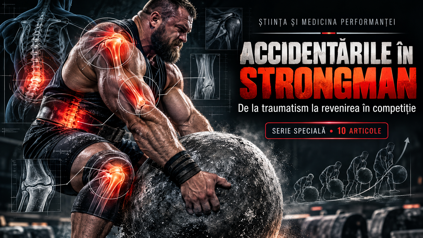
In strongman, the knee often works quietly while everyone watches the implement being lifted. During a heavy squat, it permits the descent and the drive back up. During a yoke carry, every step demands coordinated lower-limb control. In farmers walks, it contributes to acceleration, braking, and changes of direction. During atlas stones and sandbag loading, it helps lift the object from the ground, position it on the lap, and complete the final extension. In a truck pull, force generated through the hip and knee must be transmitted repeatedly into the ground. Each event exposes the same joint to a different combination of compression, tensile forces, joint moments, fatigue, and neuromuscular demands. Knee pain is therefore a relevant concern in strength sport, but it would be wrong to assume that every painful knee is damaged or that every knee capable of supporting extraordinary weight is invulnerable.
This fifth episode of “Strongman Injuries: From Trauma to Returning to Competition” examines the knee under extreme loading: the anatomy that makes force production possible, plausible injury mechanisms, patellar tendinopathy, patellofemoral pain, meniscal and ligament injuries, ruptures of the extensor mechanism, degenerative changes, and the journey from initial symptoms to competition. We will explore the meaning of pain that develops over weeks, when a pop during an event warrants stopping, why sudden joint swelling changes the assessment, and why MRI findings must be considered alongside clinical function. Exercise rehabilitation will be discussed as an important tool without confusing it with permission to continue maximal lifts regardless of symptoms.
In a retrospective study of 213 strongman athletes, Winwood and colleagues reported that the knee accounted for approximately 11% of recorded injury locations over a twelve-month period [1]. This does not mean that 11% of all strongman participants ruptured an anterior cruciate ligament, nor does it establish an 11% annual injury risk for any particular event. The researchers used retrospective self-reporting of injuries that resulted in missed or modified training sessions or competitions. Individual injuries were not all confirmed through imaging, and comparable risk rates were not established across every event. Nevertheless, the finding confirms that the knee matters to long-term athlete health, even though the lower back and shoulder often receive more attention.
One principle will return repeatedly throughout this article: mechanical loading is not synonymous with tissue injury. A healthy joint can tolerate substantial forces through the combined work of its muscles, bones, cartilage, ligaments, and training adaptations. Problems may follow acute trauma, rapid changes in exposure, irritated tissues, existing joint disease, or interactions between several factors. Causation cannot be determined from one video clip, and pain alone does not demonstrate a tear. At the same time, a new inability to actively extend the leg, visible deformity, rapid swelling, or inability to bear weight should not be dismissed because “something always hurts in strongman.”
Knee anatomy: more than a hinge between two bones
The knee contains the tibiofemoral and patellofemoral articulations. The femur meets the tibia, while the patella moves along a groove at the distal end of the femur. The fibula does not form the principal weight-bearing tibiofemoral surface, but it is important for various ligament and muscle attachments, especially laterally. Flexion and extension dominate knee movement, but the joint also allows components of rotation and translation, particularly in certain positions. This complexity explains why knee function cannot be assessed solely by asking whether an athlete can bend the leg.
Articular cartilage covers bone surfaces involved in motion and helps distribute forces alongside synovial fluid and other joint structures. The medial and lateral menisci are fibrocartilaginous structures between the femur and tibia. They contribute to load distribution, joint congruence, and stability. They are not disposable pads that can be removed without consequence. Understanding their role helps explain why modern meniscal surgery prioritises preservation of functional tissue whenever intervention is necessary and feasible [4–6]. Meniscal tears may arise after trauma, occur in a degenerative context, or be discovered without being the primary source of symptoms.
The anterior and posterior cruciate ligaments contribute to controlling the translation and rotation of the femur relative to the tibia. The medial and lateral collateral ligaments help resist forces tending to open the joint on either side. The capsule and posterolateral structures complete this system. Stability, however, is not provided by ligaments alone. The quadriceps, hamstrings, calf muscles, and hip musculature all influence dynamic load control. An athlete can have good passive stability but insufficient movement capacity for a highly demanding event. Conversely, exceptional muscular strength cannot reliably compensate for every major ligament injury.
The knee extensor mechanism includes the quadriceps, quadriceps tendon, patella, and patellar tendon attaching to the tibial tuberosity. Through this continuous structure, force generated by the quadriceps is transferred into knee extension. The patellar tendon is sometimes called the patellar ligament, but its function in the extensor mechanism should be distinguished from that of the cruciate ligaments. Pain at the front of the knee can originate from several tissues, and a complete patellar tendon rupture has profoundly different consequences from gradually developing tendinopathy. Describing pain “just below the kneecap” is not sufficient to treat these conditions as equivalent.
Other tissues receive less attention in gym conversations: the infrapatellar fat pad, bursae, synovial lining, patellar retinacula, and periarticular structures. They can contribute to painful presentations, but tenderness at a particular location does not establish a diagnosis. Swelling has very different implications when it follows an acute twisting injury, develops gradually after repeated loading, or appears with fever. Anatomy is not intended as a catalogue of frightening terminology. It is the map through which history and physical examination become meaningful.
How the knee handles large forces: compression, tension, and dynamic control
During a squat, body mass and external weight produce moments that muscles must counteract. As knee position and flexion change, the demand placed on the quadriceps and the forces at the patellofemoral and tibiofemoral articulations also change. These variations do not automatically define a movement as dangerous. Joint forces are an ordinary part of knee function, and suitable training can develop muscular capacity and functional tolerance. Injury risk cannot be inferred solely from calculated compressive forces because pain, tissue damage, anatomy, and individual capacity do not follow a simple one-to-one relationship.
A knee passing in front of the toes during a squat is not automatically committing a technical error. Artificially preventing forward knee travel may redistribute demands towards the hips, trunk, and other segments without guaranteeing injury prevention. For some athletes, a particular range or position is more comfortable; for others, deep flexion is both functional and technically necessary. There is no universally safe knee angle and another angle that inevitably causes harm. In a sport dominated by irregular objects, a rigid rule based only on knee position relative to the shoe is insufficient. Current capacity, progressive loading, movement control, and the clinical response matter more.
During a yoke carry, the task includes lifting the load from the supports, maintaining its position, and taking repeated steps under weight. The knees repeatedly control force absorption and production as the combined centre of mass shifts slightly with each step. Acceleration, braking, and correction of frame oscillation change muscular and joint demands. This offers a plausible explanation for symptoms in athletes unfamiliar with the implement or exposed to very high training volumes, but it does not prove that yoke carries cause ligament ruptures more often than other events. Strong prospective comparisons specific to strongman remain unavailable.
Farmers walks and frame carries demand knee control during movement, but the initial pick-up and the subsequent run do not necessarily distribute mechanical requirements in the same way. During a truck pull, repeated powerful pushes against the ground drive the load forwards, while trunk position, harness design, footwear, and traction alter the mechanics. Sled pushing and dragging involve resistance vectors different from axial squatting. It is unjustified to declare an event universally “good for the knee” or “bad for the meniscus” simply because it looks less dramatic. The relevant exposure includes force direction, step count, distance, speed, range of motion, and the athlete's history.
Atlas stones and sandbag loading combine knee flexion with lifting objects that may restrict freedom of movement. When bringing a stone into the lap, an athlete can create direct contact between the object and thighs; both hip and knee motion contribute to the final load. Some medleys place events in rapid sequence, leaving little time for movement control to recover. An event repeated many times should not be treated as mechanically equivalent to a single maximal attempt even if the total nominal tonnage seems similar. The knee responds to the complete exposure, not just the weight written on one implement.
Pain at the front of the knee: one region, several diagnoses
Anterior knee pain is a descriptive umbrella, not a diagnosis. An athlete may have patellofemoral pain, patellar tendinopathy, quadriceps tendon symptoms, irritation of the infrapatellar fat pad, or a problem involving the joint itself. Pain distribution, provoking activities, onset, and clinical examination help distinguish these possibilities. Gradually developing pain after an increase in squat volume does not prove that anything has ruptured. Sudden pain with loss of active knee extension, however, should not be treated as ordinary tendinitis. Similar locations can contain conditions with very different treatment pathways and prognoses.
Patellofemoral pain usually refers to discomfort around or behind the patella, aggravated by squats, stair climbing, or other activities involving loaded knee flexion. It should not automatically be equated with “worn-out kneecap cartilage,” “destroyed cartilage,” or a single alignment defect. It is multifactorial, involving load tolerance, muscle capacity, movement strategies, and individual characteristics. A 2024 best-practice guide recommends education and knee-targeted exercise as core treatment, while hip exercises and supporting interventions can be tailored to the person's particular presentation [2].
In strongman, patellofemoral symptoms may be most noticeable during squats, controlled lowering of heavy objects, or repeated lower-body training. Yet it is not justified to blame every case on a technical fault or supposedly “sleeping glutes.” Assessment should identify which tasks provoke symptoms, how long the response lasts after training, and which variations are tolerated. Temporarily changing range of motion or volume may allow preparation to continue. Other athletes may need a broader exercise programme addressing quadriceps and hip capacity. Education should replace fear and promises of instant cures with achievable, measurable functional goals.
The 2024 guideline emphasises individualisation. Someone who tolerates loaded knee flexion but demonstrates quadriceps weakness may benefit from knee-focused strengthening. Another person with highly irritable symptoms during flexion may initially require different forms of loading before gradually progressing [2]. Taping, certain prefabricated foot orthoses, or manual techniques can be considered as supporting tools when appropriate, but they are not automatic substitutes for exercise and education. In competitive sport, the ultimate goal is not merely being comfortable on stairs; it is developing the capacity to manage event-specific demands. Applying general clinical recommendations to extreme loads nevertheless requires caution.
Pain can persist even when imaging does not reveal a dramatic lesion, and imaging abnormalities may also exist without important symptoms. Neither scenario justifies panic or accusing an athlete of imagining the problem. Knee function cannot be reduced to a scan, and neither can it be measured solely by the ability to perform one heavy squat on a good day. Symptom trends over weeks, ordinary daily activities, and the response to progressive loading all matter during recovery.
Patellar tendon: from normal adaptation to tendinopathy
Patellar tendinopathy is characterised by load-related pain involving the patellar tendon, often near the lower pole of the patella. It is well known in jumping sports, but the substantial loading of the extensor mechanism in strength sport makes it relevant here as well. Not every painful spot in this region represents tendinopathy, and the popular word “tendinitis” does not accurately describe all the biological processes in chronic presentations. Persistent symptoms may involve structural changes and complex pain mechanisms that cannot be explained by conventional inflammation alone. Diagnosis is principally clinical, with ultrasound or MRI useful in selected circumstances [3].
A tendon transfers muscular force to bone. Because it is designed to tolerate loading, it can also adapt to training, although the response depends on exposure and individual history. An athlete who moves rapidly from modest squat volume to repeated heavy squats, loading events, and carries may temporarily exceed the capacity of certain tissues. This does not mean those exercises are intrinsically harmful or that tendinopathy always results from simply “doing too much.” Age, previous symptoms, recovery conditions, and the interaction between volume and intensity may all be relevant. When load-related pain appears, examining the entire programme is more useful than searching for one supposedly guilty repetition.
Tendon pain is frequently related to activity, but it does not always follow a predictable pattern. Some athletes feel discomfort during warming up, temporary improvement once training begins, and increased pain later that evening or the following morning. This pattern is not a definitive diagnostic test. Stiffness at the start of movement and pain during repeated squatting, lowering, or rising may also be reported. In contrast, sudden severe pain with a pop and a new inability to extend the knee actively suggests a different problem and warrants prompt evaluation for a possible extensor-mechanism rupture. Continuing heavy loading to “warm up the tendon” is inappropriate in that situation.
Conservative management of patellar tendinopathy emphasises load management and progressive exercise. A clinical review in the Journal of Athletic Training describes heavy slow resistance training, selected isometric exercises, and stages of sport-specific reloading according to symptom irritability [3]. It does not follow that every patient should use the same weight, repetitions, or exercise sequence. A strongman athlete may need changes in range of motion, tempo, and session frequency before returning to complex loaded events. The real objective is restoring functional capacity, not merely creating a brief window of pain relief.
In a randomised clinical trial involving 76 people with patellar tendinopathy, Breda and colleagues compared progressive tendon-loading exercises with an eccentric-focused programme and found better clinical outcomes after twenty-four weeks in the progressive-loading group [9]. This supports structured progression, but the participants were not exclusively strongman athletes squatting or carrying extreme weights. The findings cannot determine a precise implement weight at which every competitor becomes ready to return. They do, however, explain why an active, staged approach makes more sense than indefinite rest or daily maximal testing despite pain.
Tendinopathy and tendon rupture are distinct conditions. A painful tendon may remain functional, while imaging abnormalities can also be found in people with relatively few symptoms. Partial and complete tears raise different clinical questions, particularly when active extension has been compromised. Stories about athletes competing for years with an “inflamed tendon” do not establish that all anterior knee pain can be ignored. Equally, imaging changes do not automatically require surgery. The specific diagnosis and the athlete's actual functional demands must guide management.
Extensor-mechanism rupture: when this is no longer ordinary overload
The quadriceps tendon connects the quadriceps muscles to the patella, while the patellar tendon continues the system to the tibia. A complete rupture of either structure can compromise the ability to extend the knee actively or hold the leg straight against gravity. Athletes may describe a pop, sudden pain, swelling, altered contours, or the feeling that the knee has given way. Some movements may remain possible despite a serious lesion. Suspected complete extensor-mechanism rupture requires stopping the event and seeking prompt orthopaedic assessment. Depending on the tear and clinical context, surgery may be needed.
Plausible biomechanical mechanisms in strength events include large demands on the extensor mechanism during maximal attempts, unexpected eccentric loading, loss of balance, or sudden changes in limb position. Plausibility is not proof that a particular strongman event frequently causes complete tendon rupture. Diagnosis must be medically confirmed rather than declared from one replayed clip. A competitor's history, treatment options, and any postoperative rehabilitation programme cannot be replaced by generic internet advice or the judgement of someone watching the attempt from the platform.
A critical distinction exists between knee pain that still permits active extension and a new inability to straighten the knee. After a heavy attempt, an athlete may say the leg “will not support me.” The key question is not only pain intensity but whether there has been an objective loss of function. Fractures and other serious injuries can also cause substantial functional loss, so repeatedly testing the knee under weight is inappropriate. Prompt medical assessment may include examination of the extensor mechanism and selected imaging. Timing matters for certain ruptures, and postponing care to protect one competition can worsen the eventual outcome.
Meniscus: traumatic tears, degenerative changes, and incidental findings
The medial and lateral menisci distribute load between the femur and tibia. Meniscal injuries may follow flexion and rotation, sometimes producing joint-line pain, swelling, or mechanical symptoms. But pain along either side of the knee does not confirm a tear, and painless clicking alone is not a diagnosis. Assessment may include the precise injury history, movement range, tenderness, effusion, and selected clinical tests. Importantly, the AAOS guideline published in 2024 focuses on acute isolated meniscal injuries, not every degenerative tear or every tear occurring alongside a major ligament injury [4].
In strongman, the knee may be flexed while the athlete makes an unexpected directional correction, perhaps during a farmers walk turn or while recovering balance during a loading medley. This creates a plausible mechanism for stressing meniscal structures, but it does not mean every loaded twist causes a tear. Current data cannot assign a credible event-specific percentage of meniscal risk to each implement. Acute injuries may cause pain, swelling, and restricted movement, but presentations vary. A genuinely locked knee, where a mechanical obstacle prevents normal extension, needs prompt clinical assessment. Attempting to force the knee open with heavy squats is not a sensible response.
The word “locking” requires care. Severe pain can make a person reluctant to move the knee without a structure physically preventing extension. This differs from true mechanical obstruction caused by displaced tissue. The distinction can affect orthopaedic decision-making. AAOS guidance supports using clinical examination alongside imaging when appropriate, with MRI preferred for diagnosing acute isolated tears. It also emphasises preserving as much functional meniscal tissue as possible when surgery is indicated [4]. Claiming that every torn meniscus must be removed ignores the very biological function treatment aims to preserve.
Degenerative meniscal lesions exist in a different clinical context from acute traumatic tears. Some people have MRI findings that do not fully explain their symptoms and do not necessarily require arthroscopy. Decisions depend on symptoms, examination, tear configuration, true locking, accompanying disease, and personal goals. An international ESSKA–AOSSM–AASPT consensus developed in 2024 and published in 2025 identifies nonoperative treatment as an important first-line approach for degenerative lesions and a potential option for some acute tears [6]. This does not imply that every acute tear should be managed identically or that surgery has no remaining role.
Following meniscal surgery, rehabilitation differs markedly between partial meniscectomy and meniscal repair. Removing a small portion of tissue and suturing a tear are biologically and mechanically different interventions. A 2025 international consensus recommends progression based on clinical milestones after meniscectomy, while repair and reconstruction require attention to both time and functional criteria [5]. A single universal timetable would therefore be irresponsible. Returning to heavy squats or yoke carries requires more than painless walking; range of motion, effusion, muscular strength, the type of repair, and the treating team's instructions all matter.
Restoring full knee extension is an important objective in various postoperative and postinjury settings. Persistent extension loss can alter gait and task mechanics, while joint effusion may inhibit effective quadriceps activation. A strongman should therefore not assess the injured knee solely by the weight that can be moved on a leg press. Movement, control, and the response after activity are equally relevant. Where postoperative restrictions exist, the individual's surgical protocol takes precedence over a general training article, because tissue healing and the specific repair determine the acceptable progression.
Cruciate and collateral ligaments: stability cannot be measured in kilograms
The anterior cruciate ligament, or ACL, contributes to controlling forward movement of the tibia and certain rotational forces. ACL ruptures have been studied extensively in sports involving cutting, rapid changes of direction, and landing, but they can occur in other traumatic situations as well. Twisting trauma may be accompanied by a pop, rapid swelling, or a sense of giving way. These signs do not establish the diagnosis in isolation, and being able to walk immediately after the event does not exclude rupture. Suspected ACL injury warrants clinical examination, while associated meniscal or cartilage injuries may influence treatment decisions [7].
In strongman, events performed mainly in straight lines might appear to involve little rotation, but the real environment includes turns, balance corrections, uneven surfaces, and implements that can shift unexpectedly. These factors justify training dynamic knee control, but they do not establish a known incidence of ACL tears in yoke carries or farmers walks. Event-specific epidemiological data are too limited for confident estimates. Directly transferring injury rates from football or handball to strongman would be misleading. When a competitor becomes injured, the relevant questions concern the individual mechanism and examination rather than the event's reputation.
The posterior cruciate ligament, or PCL, limits posterior displacement of the tibia and may be injured following an impact to the front of the shin in certain positions, hyperflexion, or other trauma. Medial and lateral collateral ligament injuries may follow forces that open one side of the joint and can be combined with capsular or posterolateral damage. Multiligament injuries have very different implications from a mild isolated sprain. After major trauma involving deformity, marked instability, or suspected knee dislocation, delaying assessment can be dangerous. Some multiligament patterns also threaten nerves and blood vessels.
A ligament diagnosis cannot be inferred solely from the feeling that the knee is “giving way.” Perceived instability can be influenced by pain, swelling, and muscular inhibition. A qualified examination may include specific stability tests and investigation of associated lesions, with MRI used when the clinical picture warrants it. If the knee has sustained major trauma and the foot becomes pale or cold, new numbness develops, or there is obvious deformity, emergency assessment is necessary. Competing again because a tight wrap seems to improve support is not an adequate substitute for evaluation.
ACL treatment may be operative or nonoperative depending on the tear, instability, associated damage, physical demands, and informed patient preferences. The AAOS guideline published in 2022 provides recommendations for individualised decision-making and, where reconstruction is indicated, considerations concerning timing and graft choice [7]. It is too simplistic to say every ACL rupture must be operated on. It is equally unjustified to promise that very strong muscles will always compensate for a deficient ligament. For strongman, an additional concern is whether the athlete can safely manage unusual loads and dynamic event transitions, questions for which direct research remains scarce.
Rehabilitation after ligament injury must pursue more than an impressive squat number. Relevant goals include movement range, control of swelling, leg strength and endurance, task-specific motor control, symptom response, and confidence in the joint. Following ACL reconstruction, return to sport is multidimensional; simply reaching a prescribed number of postoperative months is insufficient. A scoping review found substantial variation in how time, strength testing, hop tests, and clinical examination were used as return-to-sport criteria [10]. In strongman, progression must respect the specific operation and the demands of the athlete's intended events.
Articular cartilage and osteoarthritis: beyond the myth of “destroyed knees”
Knee osteoarthritis involves the joint as a whole, rather than being explained solely by the loss of cartilage visible on imaging. Changes may involve cartilage, subchondral bone, synovium, and surrounding structures, while pain and disability do not necessarily match radiographic severity. An athlete may have imaging changes with relatively few symptoms, whereas another may have important pain requiring treatment. No single age or bodyweight threshold makes osteoarthritis inevitable for every strength athlete. Prior trauma, medical history, and multiple individual factors influence its course.
The assertion that each heavy squat “grinds away cartilage” is too mechanical to explain a complex biological condition. Mechanical loading has important roles in musculoskeletal function, while inactivity can worsen many health problems. This does not mean every possible intensity or training volume is risk-free. More useful questions concern the individual's tolerance, injury history, strength development, and symptom response. Compression alone cannot demonstrate that osteoarthritis is occurring, and the ability to tolerate heavy lifting cannot guarantee that degenerative changes will never arise.
NICE guidance published in 2022 places tailored therapeutic exercise, education, and appropriate weight management at the centre of osteoarthritis care [8]. Local muscular strengthening and aerobic activity can improve function, even if some people experience temporary discomfort when beginning a programme. Medication and procedures require separate evaluation of benefits and risks. The idea that an osteoarthritic joint must remain unused so that it cannot “wear out” does not reflect contemporary guidance for most people. In strongman, programmes may be adapted to preserve physical activity while taking the clinical situation seriously.
Body mass matters, but should not be discussed through simplistic moral judgements. Open-class athletes can have very large bodies, high muscular development, and competitive demands unlike those of recreational exercisers. For a person with osteoarthritis and clinically relevant excess weight, reducing weight can improve pain and function, as described in guidelines [8]. That does not mean every large strongman should target the same bodyweight or that weight loss resolves all joint symptoms. Assessment should consider body composition, sporting goals, metabolic health, and individual risks without treating muscle mass itself as disease.
Another myth is that every click or crack indicates destroyed cartilage. Popping, crepitus, and other joint sounds can occur without clinically meaningful injury. Their importance depends on accompanying pain, swelling, locking, trauma, and loss of function. A knee that clicks during squats but does not hurt, swell, or lose function is not automatically seriously damaged. A pop during trauma followed by rapid swelling and instability belongs in a very different category. What accompanies the sound often matters much more than the sound itself.
Red flags: the knee that requires assessment, not another warm-up
Some presentations require immediate discontinuation of the event and urgent medical attention. Obvious deformity after injury, inability to bear weight, suspected fracture, a cold or pale foot, new numbness, or major weakness should not be managed by simply wrapping the knee and completing the contest. Suspected knee dislocation is especially serious because vascular damage can be present even if the joint appears to have spontaneously returned to normal alignment. This situation requires emergency care rather than a return to the training platform.
A very hot, swollen, painful knee, especially with fever or general illness, may suggest joint infection or another acute inflammatory condition requiring urgent assessment. Fever is not present in every case, and inability to bear weight should not automatically be attributed to a sporting injury. Rapid swelling after significant trauma may reflect bleeding into the joint or structural damage. These possibilities cannot always be distinguished without medical examination, but it is clear that waiting casually for the next training session is inappropriate when serious warning signs are present.
A new inability to actively straighten the knee or lift the leg while keeping it straight raises concern for extensor-mechanism injury among other diagnoses. True mechanical locking, when extension is obstructed by something within the joint, also warrants rapid assessment. Athletes accustomed to discomfort may minimise these symptoms to avoid withdrawing from a competition. Yet the difference between tolerable pain and objective loss of function is fundamental. Disruption of a tendon or a displaced intra-articular lesion cannot be treated by determination alone.
Unilateral calf pain and swelling after prolonged travel or immobilisation, particularly with warmth, redness, or tenderness, may raise concern for deep-vein thrombosis. Sudden breathlessness, chest pain, or fainting alongside such symptoms requires emergency medical help. These are not typical meniscal symptoms, but they deserve mention in a sport whose athletes may spend many hours in vehicles and mistake discomfort after travel for muscular fatigue. Suspected thrombosis requires medical investigation, not vigorous massage or another training session.
Clinical assessment, X-ray, ultrasound, and MRI
History-taking should begin with what happened: was there a twist, collision, fall, direct impact, or gradual onset of pain? The clinician asks when swelling appeared, whether the knee locks or gives way, where pain is located, and whether the athlete can bear weight. Previous surgeries, ligament damage, tendon symptoms, and inflammatory disorders can change interpretation. In strongman, the actual events performed, recent volume changes, equipment, and circumstances of symptom onset also matter. Calling something an “injury” is not a substitute for this chronology.
Clinical examination may address gait, active and passive movement, swelling, tenderness, ligament stability, and function of the extensor mechanism. Tests for meniscus and ligament problems can guide diagnosis, but their value is greater when interpreted together rather than as isolated tricks [4]. Severe pain or swelling may make some tests difficult immediately after trauma. Repeated aggressive manoeuvres by unqualified people are inappropriate. Medical assessment determines whether observation is reasonable or whether imaging and specialist review are required.
Following acute trauma with focal bony tenderness, significant swelling, or inability to bear weight, plain radiography is commonly an appropriate initial imaging test, consistent with ACR criteria [11]. Clinical tools such as the Ottawa Knee Rule can support selected decisions about radiographs but do not replace medical judgement. Ultrasound may help assess certain tendons and fluid collections. MRI provides information about menisci, ligaments, cartilage, and other soft tissues. The choice depends on the suspected problem and whether the result would change treatment. Not every knee ache after squatting requires MRI, and not every traumatic injury with a normal X-ray excludes soft-tissue damage.
MRI offers anatomical detail but cannot independently decide whether someone may safely compete in a strongman event. A meniscal finding may be incidental, a clearly ruptured ligament may require specific management, and an effusion can accompany several different disorders. Interpretation should assess agreement between the scan, symptoms, examination, and sporting demands. Functional testing is equally relevant to eventual return, although no test guarantees that another injury will not occur. Good sports medicine avoids both unquestioning faith in MRI and dismissal of investigations that could materially change care.
Some athletes understandably seek imaging for reassurance, but a scan without a clear clinical question can reveal uncertain findings and increase worry. Conversely, refusing imaging when a knee is mechanically locked, markedly unstable, or suspected of having a major tear can be inappropriate. Useful discussions explain why a particular investigation is or is not needed, what information it can provide, and how possible results might change management. The goal is not to collect images. It is to make better clinical decisions.
Medical recovery and therapeutic training: bringing two objectives together
Recovery from knee pain begins by identifying what may still be loaded and what needs protection. In patellofemoral pain or tendinopathy without evidence of rupture, progressive exercise is often central to management [2,3,9]. Following meniscal repair, ligament reconstruction, or tendon rupture, the restrictions and progression can be substantially different and must reflect the specific clinical or surgical situation. These presentations cannot all be treated through one generic protocol called “knee rehabilitation.” An early stage may address swelling and restoration of extension; another develops strength; later stages gradually reintroduce the demands of sport. The sequence and duration must be individualised.
A knee that does not hurt at rest is not automatically ready to support several hundred kilograms in competition. Temporary pain relief after massage or another intervention does not prove tendon healing either. Functional rehabilitation should consider gait, motion, active extension, quadriceps and hamstring strength, lower-limb control, and the response to increasing external loads. When substantial structural injury exists, the clinician also establishes relevant restrictions. Reintroducing maximal squats because an athlete feels good on a particular afternoon may disregard tissue-healing processes that are not externally visible.
Isometric exercises, slower dynamic movements, unilateral variations, and resistance training can serve different purposes. An isometric contraction may provide controlled initial loading, but it cannot automatically replace the ability to decelerate a moving yoke. A tolerable leg press can support muscular strength without recreating a farmers walk. Light squatting can be an important stage, yet rehabilitation cannot end there when the athlete intends to return to strongman. Progression should ultimately reflect positions, speed, repetitions, duration, and disturbances resembling the sport, provided the diagnosis and clinical condition allow.
For certain musculoskeletal conditions, limited and temporary discomfort during supervised exercise can be compatible with rehabilitation. There is, however, no universal pain threshold that authorises every load or every injury. A partially torn tendon, repaired meniscus, fracture, and severely unstable joint require different restrictions. Useful criteria include avoiding persistent deterioration, maintaining or improving function, observing symptoms over the next day or two, and following the treating specialist's plan. New swelling, locking, or loss of active extension warrants reassessment rather than a more aggressive warm-up.
Returning to events: squats, yokes, farmers walks, truck pulls, and loading
For squats, rehabilitation may begin with variations that allow controlled range and intensity, move towards more demanding sets, and only later approach competitive requirements. Choosing between high-bar, low-bar, front, box, or machine-supported squats should not depend on universal claims that one style is inherently safe for every injured knee. Each changes aspects of leverage and muscular demand. The choice depends on symptoms, experience, and training goals. Weight, depth, and repetition count do not all need to increase simultaneously. Temporarily reducing one variable can preserve consistency when the current progression is poorly tolerated.
Yoke carries have at least two distinct functional demands: lifting the implement and moving with it. An athlete may tolerate picking up the frame but develop pain after dozens of steps. Another may tolerate lighter walking yet remain unprepared for a near-maximal pick. Separating these variables makes responses easier to interpret. Static holds, controlled steps, distance, turns, acceleration, and eventually high-intensity efforts can be reintroduced as clinically appropriate. This illustrative sequence does not replace postoperative restrictions or the assessment of significant instability.
Farmers walks and frame carries also involve grip and trunk control, but the knees must handle acceleration, braking, implement oscillation, and turns. A straight ten-metre course does not reproduce every demand of a competition that includes tight turns or obstacles. Rehabilitation should therefore rebuild not only the ability to carry a certain weight but also the ability to remain coordinated under fatigue. Event transitions can be introduced gradually without attempting an entire competition medley during the first return-to-training sessions.
Truck pulls and sled work are often used as alternative ways of training leg drive, but their suitability depends on their real demands. Resistance, trunk angle, surface, distance, and footwear change the mechanics. A sled drag may be tolerated during a phase when deep squatting causes pain, but that does not make it a universal treatment for the patellar tendon or meniscus. An extremely demanding truck pull may be entirely inappropriate early after injury. Each variation is justified by how it serves an identified goal and by the individual's clinical response.
Atlas stones and sandbag loading should be reintroduced with the separate phases in mind: approaching the object, gripping it, bringing it to the lap, repositioning, and completing the final extension. Some stages may be tolerated better than others, and repeated loading can matter more than a single light attempt. Being comfortable with a conventional deadlift does not automatically prepare someone to lift ten stones against the clock. Rehabilitation must account for implement geometry and event pace, without assuming that general leg strength transfers completely to every task.
Knee sleeves, wraps, shoes, and medication: help versus false reassurance
Neoprene knee sleeves may provide warmth, comfort, and a subjective feeling of support. Tight knee wraps used in some strength disciplines change the mechanical conditions of a squat and can affect performance. Neither is equivalent to repairing a torn ligament or restoring a damaged meniscus. Feeling less pain while using equipment does not prove that a structural injury has healed. In selected medical conditions, a prescribed orthosis serves a different purpose and should be used according to its specific instructions.
Footwear influences foot position and contact with the ground. An elevated heel can change movement geometry in squatting, while other shoes may be more suitable for yokes or truck pulls. No shoe design, however, can guarantee prevention of knee injuries. Selection should consider the event, traction, competition rules, and individual comfort. Abruptly changing footwear before a major contest may alter movement habits; familiarisation is more useful than hoping for an instant technological cure.
Analgesics and anti-inflammatory medicines may have selected clinical uses, but their contraindications and risks matter. Masking pain before a maximal event is not the same as healing. Injections, commercially promoted regenerative procedures, and supplements advertised as cartilage-rebuilding solutions should not be presented as universal cures. Evidence varies substantially between interventions and diagnoses, and some are suitable only in carefully defined situations. For osteoarthritis, NICE places exercise and appropriate weight management at the centre of care, with medication used cautiously when necessary [8].
Prevention: managing load without promising zero risk
Prevention in strongman cannot mean eliminating squats or knee flexion. Competition demands flexion, extension, stabilisation, and loaded movement; banning those functions would remove much of the sport. A more useful approach develops muscular capacity, coordination, and familiarity with implements while progressing intensity and volume. Yet even good programming cannot guarantee that tears or painful episodes will never occur. Individual factors and unpredictable trauma remain relevant, and strongman-specific literature cannot yet quantify the benefit of every proposed preventive intervention.
When planning a training week, it is worth examining how heavy squats overlap with yokes, farmers walks, loaded accelerations, and loading events. The combined burden cannot be expressed completely by implement weight because time under tension, distance, step count, and changes of direction also matter. Someone may tolerate two events individually but not both performed at maximum intensity in the same session. This possibility should be explored through individual monitoring rather than treated as an inflexible law. The aim is enough exposure to stimulate adaptation while allowing genuine opportunities for recovery.
Competition preparation introduces additional uncertainty: travel, altered schedules, unfamiliar equipment, different surfaces, and implements chosen by organisers. An athlete who rehearses only a perfect straight yoke run may struggle when turning or braking in a confined area. Someone who trains only controlled squats may be surprised by the explosive requirements of another event. Specific training should expose the athlete progressively to such challenges within present capacity. Competition day is not the ideal occasion for first contact with the basic technique of an unfamiliar implement.
Sleep, general fatigue, health conditions, and occupational demands can influence preparedness, but they should not become simplistic explanations for every injury. One athlete can have inadequate sleep without injury, while another can be hurt after excellent recovery. Monitoring is meant to reveal useful patterns and support decisions, not predict the precise timing of the next trauma. If pain worsens consistently, swelling follows every session, and performance declines, automatically adding weight is not an intelligent strategy.
Technique in video: knee movement is not a diagnosis
During a heavy squat, knees do not necessarily move along perfectly vertical or identical paths on every repetition. Their positions reflect body structure, stance width, foot rotation, hip mobility, and the selected load. Inward knee movement, frequently called “valgus” in gym conversations, is part of a three-dimensional phenomenon that cannot be measured accurately from a single frontal video. It may alter how effort is distributed and may be relevant to coaching, but seeing it does not establish ligament rupture, meniscal damage, or a measurable individual injury risk. Near maximal loads, changes in movement strategy must be interpreted in context.
One athlete performing a maximal squat may show a modest change in knee position without pain or later symptoms. Another may perform visually orderly repetitions yet develop gradually worsening pain. These examples illustrate why technique and diagnosis are different things. Technique deserves attention for efficiency, control, and repeatability, but video analysis cannot establish the integrity of every ligament. When new instability, swelling, or functional loss occurs, clinical assessment should take priority over debate about the knee angle captured in one frame.
Stance width and squat depth can change the relative demands on the hips and knees, but strongman-specific evidence does not establish one universally protective stance. The best option depends on the athlete's goals, body proportions, training experience, and current tolerance. For a symptomatic knee, changing movement angles may be a useful method of adjusting the dose of exercise, not proof that the previous variation was inherently wrong. Controlled variety may help develop tolerance to multiple positions, particularly because strongman events do not always reproduce an identical stance.
Athletes are sometimes told to push the knees as far out as possible on every squat. Applied indiscriminately, this instruction may create unwanted compensations or force an unnatural posture. A coaching cue is useful when it improves an individual's control and performance, not when it makes them fearful of every small movement variation. When pain or instability develops, coach and athlete can review range, trajectory, and loading while seeking medical assessment when indicated. The message is not that technique is irrelevant, but that it requires careful interpretation.
Monitoring during a season: what symptoms may reveal before a major setback
Some injuries occur suddenly without useful warning signs, and no monitoring method can predict every rupture. Other problems develop gradually. An athlete may notice that the usual warm-up causes increasing discomfort, swelling persists after sessions, or an event previously tolerated now requires much longer recovery. These observations are not diagnoses, but they can indicate a need to reassess training and clinical status. A simple diary may document pain, its circumstances, swelling, movement tolerance, and effects on daily activities without pretending that subjective symptoms can be converted into perfectly objective numbers.
The distinction between an isolated fluctuation and a persistent trend matters. Mild discomfort after an unfamiliar session is not automatic proof of structural injury. However, repeated worsening, loss of full extension, or swelling that consistently returns after exercise requires attention. Monitoring should include periods when competitors modify training or miss events, not just successful personal records. Focusing only on sporting performance can hide declining function, particularly when an athlete begins compensating or unconsciously avoiding certain movements.
One useful approach is to compare responses to familiar, submaximal tasks. An athlete can observe how the knee behaves during a light squat variation, stair climbing, or a few steps with a manageable implement. Daily maximal testing is unnecessary to assess whether rehabilitation is progressing. Consistent function and an absence of worsening can be more informative than a one-off display of strength. When medical restrictions apply, such activities may need modification or postponement; they are not substitutes for professional assessment.
During the off-season, athletes have opportunities to develop tolerance to varied loads without immediate competition pressure. This may include general muscular preparation, familiarisation with implements, and carefully restored training volume. Closer to competition, specificity increases, but symptom monitoring should not disappear. A knee that swells after every maximal session deserves attention even when impressive lifts remain possible. The ability to compete today does not automatically establish that the current programme is sustainable over the coming months.
Research questions strongman has not yet answered
The retrospective work by Winwood and colleagues provides an important starting point for discussing strongman injuries, but it cannot generate precise incidence estimates for every knee disorder [1]. Comparing meniscal tear risk between squats and yoke carries, for example, would require large prospective athlete cohorts, accurate recording of event exposure, consistent diagnostic criteria, and enough medically confirmed injuries to support comparisons. Without such information, a widely circulated accident can appear representative of a much rarer phenomenon. Conversely, athletes who abandon training because of pain may disappear from datasets that include only major competition finalists.
Implement variability creates another challenge. Yokes differ in height, geometry, rigidity, and load distribution. Farmers walks may involve turns or straight courses, and competition surfaces differ in traction. The event label alone therefore describes an incomplete mechanical stimulus. Researchers should record distance, repetitions, duration, speed, surface conditions, equipment, and athlete experience alongside implement name and weight. Only then can relationships between loading and injury be examined with greater credibility.
Findings from research on patellofemoral pain, tendinopathy, and ligament treatment are valuable, but they do not represent direct trials involving elite strongman athletes [2–10]. An exercise that improves outcomes in a clinical population may be a useful rehabilitation component without proving readiness for a maximal yoke carry. Future work could examine how strength athletes progress from submaximal capacity towards competitive exposure and which functional criteria best match irregular-object events. Until then, professionals should distinguish well-supported general recommendations from necessary but incompletely tested extrapolations.
There is also an editorial problem: “risk,” “association,” and “cause” are often used as though they mean the same thing. Finding that many athletes with knee pain perform squats does not prove squats are the sole cause. Finding that the knee experiences high compression does not prove that compression is producing disease. Observational comparisons can identify associations; causal conclusions require stronger methods and attention to confounding factors. Readers interested in the science of performance gain more from these distinctions than from memorable but poorly justified verdicts.
Athlete, clinician, and coach: shared goals and different responsibilities
Following injury, competitors understandably want an exact date when they can return. Clinicians focus on diagnosis, health, and healing, while coaches focus on rebuilding performance. These perspectives should complement each other rather than compete. An international consensus describes return to sport as a continuum: return to participation, return to the sport itself, and return to previous or higher performance [12]. An athlete may achieve the first stage without being ready for a complete strongman competition, and a successful training session does not prove that the final stage has been reached.
Useful communication requires more than asking whether pain remains. The team can agree which symptoms warrant stopping, how function will be assessed, which event demands will be reintroduced, and who makes decisions when warning signs appear. After surgery, the surgeon's specific restrictions are central to programme design. If symptoms fail to improve as expected, the strategy should be reviewed before loading is increased. Such a process does not eliminate sporting risk, but it makes uncertainty more visible and decisions more accountable.
Finally, the psychological dimension of returning to sport must be recognised. Someone with an injury history may hesitate while descending into a squat, avoid turning under a carry, or interpret every sensation as another tear. That fear does not mean the condition is “all in their head.” Confidence can be rebuilt through accurate information, realistic progression, and repeated experiences of well-tolerated movement. When fear substantially limits participation, specialised psychological support may also help. Sustainable performance belongs to an athlete who understands both the strength available and the circumstances in which it is sensible to use it.
Three practical scenarios that require different responses
An athlete develops pain at the front of the knee after several weeks of increased squat volume, without a specific injury, mechanical locking, or difficulty walking. This description does not permit a remote diagnosis. Persistent symptoms may justify clinical assessment, training-load review, and an appropriate exercise programme. Patellofemoral pain and tendon pain can share features, but their treatment reasoning is not identical [2,3]. The athlete should not automatically be told that a meniscus has torn, nor should they be encouraged to continue maximal attempts regardless of symptoms.
Another competitor twists a knee during a farmers walk turn, hears a pop, and develops rapid swelling. Walking may remain possible, but instability or difficulty fully straightening the leg emerges. This situation requires stopping the event and prompt assessment for possible ligament, meniscal, bone, or combined injury. The sound alone cannot diagnose an ACL tear, and an improvised squat test cannot confirm joint integrity. Treatment decisions follow the diagnosis rather than preceding it.
A third athlete resumes training after meniscal surgery and can tolerate moderate leg-press weights. That does not establish readiness for maximal yokes or a long loading medley. If the meniscus has been repaired, the biological and rehabilitation considerations differ from those following partial meniscectomy [5,6]. Surgical clearance, control of swelling, restored strength, and progressive event-specific testing matter more than the excitement of the first pain-free workout. The goal is a sustainable season, not a single demonstration of determination.
Conclusion: a strong knee is a trained system, not an indestructible part
A strongman athlete's knee can withstand extraordinary demands, but function depends on multiple structures working together. Cartilage, menisci, ligaments, the patellar and quadriceps tendons, muscles, and the nervous system contribute in different ways to performance. Pain may exist without a major rupture, while certain symptoms require prompt assessment for serious pathology. A mature approach rejects both extremes: that every deep squat destroys the joint and that every symptom can be solved with another heavy set.
Returning to competition requires more than the disappearance of pain. It means recovering movement, strength, control, and tolerance for the exact mechanical demands of the chosen events, while respecting tissue healing when a structure has been repaired. Available evidence supports useful principles, not universal formulas for extreme loads. Protecting the long-term ability to compete requires appropriate diagnosis, thoughtful progression, medical collaboration, and respect for genuine warning signs. A strong knee is not one that has been forbidden to move, but one that can meet sporting demands without important symptoms being ignored.
References and bibliography
[1] Winwood PW, Hume PA, Cronin JB, Keogh JWL. Retrospective injury epidemiology of strongman athletes. Journal of Strength and Conditioning Research. 2014;28(1):28–42. doi:10.1519/JSC.0b013e3182986c0c. https://pubmed.ncbi.nlm.nih.gov/23669816/
[2] Neal BS, Lack SD, Bartholomew C, Morrissey D. Best practice guide for patellofemoral pain based on synthesis of a systematic review, the patient voice and expert clinical reasoning. British Journal of Sports Medicine. 2024;58:1486–1495. doi:10.1136/bjsports-2024-108110. https://bjsm.bmj.com/content/58/24/1486
[3] Rosen AB, Wellsandt E, Nicola M et al. Clinical Management of Patellar Tendinopathy. Journal of Athletic Training. 2022;57(7):621–631. https://meridian.allenpress.com/jat/article/57/7/621/471608/Clinical-Management-of-Patellar-Tendinopathy
[4] American Academy of Orthopaedic Surgeons (AAOS). Management of Acute Isolated Meniscal Pathology: Evidence-Based Clinical Practice Guideline. June 10, 2024. https://www.aaos.org/quality/quality-programs/acute-isolated-meniscal-pathology/
[5] Pujol N et al. The formal EU–US Meniscus Rehabilitation 2024 Consensus: An ESSKA–AOSSM–AASPT initiative. Part I: Rehabilitation management after meniscus surgery. Knee Surgery, Sports Traumatology, Arthroscopy. 2025. doi:10.1002/ksa.12674. https://doi.org/10.1002/ksa.12674
[6] Prill R et al. The formal EU–US Meniscus Rehabilitation 2024 Consensus: An ESSKA–AOSSM–AASPT initiative. Part II: Prevention, non-operative treatment and return to sport. Knee Surgery, Sports Traumatology, Arthroscopy. 2025. doi:10.1002/ksa.12689. https://doi.org/10.1002/ksa.12689
[7] American Academy of Orthopaedic Surgeons (AAOS). Management of Anterior Cruciate Ligament Injuries: Clinical Practice Guideline. August 22, 2022. https://www.aaos.org/quality/quality-programs/anterior-cruciate-ligament-injuries/
[8] National Institute for Health and Care Excellence (NICE). Osteoarthritis in over 16s: diagnosis and management. Guideline NG226. 2022. https://www.nice.org.uk/guidance/ng226/chapter/recommendations
[9] Breda SJ et al. Effectiveness of progressive tendon-loading exercise therapy in patients with patellar tendinopathy: a randomised clinical trial. British Journal of Sports Medicine. 2021;55:501–509. doi:10.1136/bjsports-2020-103403. https://pubmed.ncbi.nlm.nih.gov/33219115/
[10] Burgi CR et al. Which criteria are used to clear patients to return to sport after primary ACL reconstruction? A scoping review. British Journal of Sports Medicine. 2019. doi:10.1136/bjsports-2018-099982. https://bjsm.bmj.com/content/53/18/1154
[11] American College of Radiology (ACR). ACR Appropriateness Criteria: Acute Trauma to the Knee. Diagnostic imaging appropriateness guidance. https://acsearch.acr.org/docs/69419/Narrative/
[12] Ardern CL et al. 2016 Consensus statement on return to sport from the First World Congress in Sports Physical Therapy, Bern. British Journal of Sports Medicine. 2016;50:853–864. doi:10.1136/bjsports-2016-096278. https://bjsm.bmj.com/content/50/14/853
Editorial and medical note: source [1] reports retrospective strongman-specific data, with important limits for estimating the risk of individual events. Sources [2]–[12] include clinical guidelines, research, a randomised trial, and international consensus statements developed in sporting or clinical populations that are not identical to elite strongman athletes. Applying these principles to extreme loads therefore requires individual judgement; the recommendations are not validated protocols for every implement. Scenarios are hypothetical and are not accounts of real people. The material is educational and cannot replace medical assessment. Deformity, vascular warning signs, a hot and severely swollen joint, loss of active knee extension, or inability to bear weight following trauma requires prompt, sometimes emergency, evaluation.
More From The Journal
Training
The Science of Strongman Training - Episode X: Competition Programming
How to build a complete Strongman competition preparation block: calendar, events, volume, intensity, specificity, simulations, autoregulation, tapering and fatigue management.
Training
The Science of Strongman Training - Episode IX: Peaking
Peaking in Strongman: how to turn months of training into maximal competition performance by reducing fatigue, preserving adaptations and managing specificity.