
Sports Medicine
Series: STRONGMAN INJURIES: FROM TRAUMA TO RETURNING TO COMPETITION - Episode 4: The Lumbar Spine: Overload, Pain and Pathology
October 12, 2026

In strongman, the lumbar spine is not simply a segment that must be “kept straight” while the rest of the body moves the weight. It is a living structure that must transmit force, withstand compression, control motion, and participate in tasks where the implements are far less predictable than a barbell. An athlete may perform a heavy deadlift, lift an atlas stone, carry a yoke weighing several hundred kilograms, and load sandbags onto a platform in the same training week. Each event challenges the lower back through a different combination of force, fatigue, posture, and speed. Pain arising in this environment is not automatic proof that the spine has been damaged, but it should not be dismissed as an unavoidable price of performance. Between trivialising every symptom and catastrophising every sensation lies a more useful approach: clinical assessment, an understanding of mechanisms, and intelligent management of loading.
This fourth episode of “Strongman Injuries: From Trauma to Returning to Competition” examines the lumbar spine through the lenses of anatomy, overload, acute and persistent pain, disorders requiring medical attention, and criteria for responsible return. Deadlifts, yoke carries, farmers walks, atlas stones, tyre flips, Zercher carries, and loading medleys will appear throughout, but an important distinction must be maintained: the fact that an event generates high mechanical forces does not, by itself, demonstrate that it causes a particular injury. We will separate plausible biomechanical mechanisms from clinical observations and conclusions supported by research. This matters particularly in strongman, where the epidemiological literature remains limited compared with research involving the general population, team sports, or more established strength disciplines.
The importance of the lower back is supported by data, not merely by impressions in the gym. In a retrospective survey involving 213 strongman athletes, Winwood and colleagues reported that the lower back accounted for approximately 24% of reported injury locations and was among the most frequently affected regions [1]. Across the same twelve-month period, 82% of participants reported at least one injury. These numbers must be interpreted precisely. They do not mean that 24% of strongman participants have a herniated disc or that one quarter of lifts cause lumbar injury. The study relied on retrospective self-reporting, and the injury definition required a missed or modified training session or competition. Its findings identify an important problem without establishing individual imaging diagnoses or the causal role of any specific strongman event.
There is another reason to avoid simplistic explanations. For a strength athlete, the back is not merely a body region but part of the physical system on which competitive identity depends. Pain that is minor during daily life may become decisive when stabilising a yoke, accelerating a barbell from the floor, or controlling a shifting sandbag. Conversely, an athlete with extraordinary strength may show substantial imaging changes without clinically important symptoms. A personal record, the appearance of an MRI scan, and pain intensity considered separately cannot explain the entire picture. Sports medicine becomes useful when it combines personal history, clinical examination, symptom distribution, athletic goals, and the functional consequences of the condition.
Lumbar anatomy: the architecture that transfers force
The lumbar region usually consists of five vertebrae, L1 through L5, connected by intervertebral discs, posterior joints, ligaments, and an elaborate muscular system. Discs are not soft cushions that simply “slip” whenever a heavy load is lifted. They contain a nucleus pulposus and annulus fibrosus with biological and mechanical properties involved in force transmission. Vertebral bodies, endplates, and facet joints help distribute loading. Nerve structures pass through openings between vertebrae, while the spinal canal contains neural structures continuing towards the legs. In the lower lumbar region, the spinal cord typically has already ended at a higher level, and the nerve roots form the cauda equina. This distinction is especially important when discussing serious neurological warning signs.
The erector spinae, multifidus, abdominal muscles, diaphragm, pelvic floor, and hip muscles all contribute to trunk control. They do not operate as isolated components activated perfectly by one magical exercise. Stability emerges from coordinated action in response to load, position, speed, and task. During a deadlift, hip and knee extension produce much of the movement while the trunk muscles control positioning and force transfer. During atlas stone loading, the trunk may contribute more visibly as the athlete moves around the implement. Motion at a spinal segment or fatigue in the back muscles does not automatically mean injury. Range of motion, repetitions, prior preparation, current symptoms, and clinical findings all matter.
Axial compression, shear forces, and flexion or extension moments describe different components of mechanical loading. Compression presses structures together, shear describes a tendency for relative sliding, and a flexion moment describes the tendency of an external load to rotate the torso forwards. In an actual event, these forces coexist and change continuously. A barbell held close to the body may create different lever arms from a bulky stone, but this observation cannot be turned into a universal ranking of dangerous exercises. Absolute weight is only one variable. The distance between the load and the body, the duration of muscular effort, changes of direction, and the athlete's capacity on that particular day also shape the task.
Mechanical adaptation underpins strength training. Bone, muscle, and connective tissue respond to loading, although their biological mechanisms and adaptation rates differ. The spine does not become inherently fragile merely because it is used intensively, yet the ability to tolerate specific demands varies between individuals and across training phases. Someone with years of progressive exposure may manage training sessions that would overwhelm an inexperienced lifter. Equally, a history of impressive lifts does not grant immunity from fatigue, abrupt volume changes, trauma, or illness. Good programming is not about eliminating force. It is about improving the athlete's capacity to manage it.
What a “lumbar injury” means, and what it does not
In gym conversations, almost any lower-back pain receives one of two improvised diagnoses: “muscle spasm” or “herniated disc.” The reality is much more varied. Pain may be non-specific, muscular, related to the posterior spinal joints, disc-associated, referred from the hip, associated with the sacroiliac region, due to a bone injury, or connected with irritation of a nerve root. Sometimes a specific disorder can be identified. In many episodes, the available examination cannot reliably assign the entire pain experience to a single anatomical structure. “Non-specific” does not mean imaginary or unimportant; it means that a precise structural cause has not been demonstrated. The distinction helps avoid inappropriate restrictions and treatment claims that exceed the evidence [2,6].
Acute low-back pain may begin during one repetition or several hours after a demanding session. An onset during deadlifting does not prove that a disc was injured at that very moment. Tissue sensitivity, protective muscle responses, local irritation, or an existing condition becoming symptomatic can produce overlapping presentations. “I felt a sharp pain” describes an experience, not a confirmed diagnosis. Some athletes remember the exact movement, while others develop symptoms through an accumulation of exposures that cannot be assigned to a single instant. History-taking therefore examines the evolution of pain, whether it travels, which activities influence it, neurological function, previous episodes, and recent changes in training.
Chronic low-back pain, often defined in clinical research as persisting beyond about twelve weeks, does not necessarily mean tissue is continuing to tear. Over time, the sensitivity of the nervous system, sleep, stress, expectations about sport, avoidance behaviours, and physical conditioning can affect the pain experience. These influences do not remove its biological components and do not make the condition “only psychological.” A biopsychosocial perspective seeks to explain why two people with similar structural findings may experience profoundly different pain and disability. Understanding that variability can support rehabilitation, but it must never be used to dismiss symptoms or bypass investigation of serious warning signs [7,8].
“Inflammation” is another word commonly used too loosely. Training can produce normal biological responses, but not every painful sensation signals pathological inflammation or requires anti-inflammatory medication. Some symptoms improve after a temporary adjustment in training load; others respond to better sleep or require specific diagnostic investigation. Pain intensity is also an imperfect measure of structural damage. Serious disorders may initially produce subtle symptoms, while intensely painful episodes can occur without major identifiable damage. Decisions should therefore not be based solely on a numerical pain rating or a dramatic description.
Maps of loading: deadlifts, yokes, and irregular objects
The deadlift is the event most immediately associated with lumbar injury, but it is a coordinated effort involving legs, hips, trunk, and arms. Technique changes with body proportions, bar height, equipment, and competition rules. A conventional pull from the floor is not mechanically identical to an axle deadlift, block pull, elevated implement, or timed repetition challenge. An athlete might tolerate controlled training sets but develop symptoms during a medley involving faster transitions, heavier breathing, and repeated manipulation of different loads. Fatigue, accumulated work, and changing task demands may be relevant. The explanation cannot always be reduced to a single visual technical fault.
Before a maximal deadlift, an athlete sets the bar, develops tension, distributes pressure through the feet, and coordinates hip and knee extension. Bracing, the coordinated activity of the trunk muscles together with intra-abdominal pressure, is a useful strategy for many lifters. It is not, however, an absolute shield against injury or a substitute for sensible loading. Excellent bracing cannot neutralise the consequences of repeated near-maximal attempts after inadequate rest. Equally, an athlete can display apparently orderly technique and still develop persistent back pain. Biomechanics matters, but it does not explain the entire clinical picture on its own.
A yoke carry is different because the demand continues long after the implement leaves the floor. There is a pick-up phase followed by repeated steps, oscillations, corrections, and braking. Frame position, load distribution, walking speed, equipment rigidity, and surface characteristics alter the control required. When the yoke starts to sway, the trunk must react to disturbances, while fatigue can change gait and breathing strategy. Without good comparative research, it would be unjustified to claim that yoke carries cause more disc herniations than deadlifts. We can reasonably describe their characteristic combination of compression, stabilisation, and dynamic perturbation, which makes progressive exposure and familiarity with the implement important.
Farmers walks and frame carries introduce additional demands involving grip, traction, and resistance to sideways movement. Implements may swing, contact the body, or change the external moment during acceleration. If one hand loses control before the other, the athlete's movement strategy changes suddenly. This does not mean that each step with a heavy implement is wearing down the spine. It means that meaningful loading cannot be expressed in kilograms alone. Time under tension, distance, speed, and the way each turn is executed all contribute. A programme that increases weight, distance, speed, and the number of runs at once may become difficult to tolerate even for an experienced competitor.
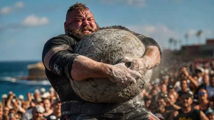
In atlas stone and sandbag loading, implement geometry matters enormously. The athlete must approach the object, bring it into the lap, reposition it, and then complete the load using coordinated hip and trunk movement. A sandbag's contents may shift and require continuous adjustment. These tasks often involve visible trunk rounding or flexion that looks different from the posture used in a conventional deadlift. Not every visible degree of spinal flexion is proof of injury, and not every final extension is dangerous. The relevant context includes load, dose, task-specific technique, training history, and symptoms; a single photograph cannot reveal the full interaction.
Tyre flipping, Zercher carries, and events with objects positioned in front of the body change the demands again. The initial drive against a tyre is different from pulling a barbell, while lifting a bulky implement changes the centre of mass of the athlete–object system. Zercher carries position the load anteriorly and change the external moment and trunk-stabilising demand. During loading medleys, transitions may matter as much as a single maximal attempt: sprinting towards the next implement, grabbing it quickly, and changing the type of effort all happen while fatigued. Training should introduce these demands progressively rather than expose the athlete to them for the first time on competition day.
Lumbar flexion: beyond dogma, towards context and evidence
Few lifting topics attract stronger opinions than the instruction to “keep your back straight.” Yet a distinction must be made between a relatively neutral lumbar posture, the whole torso leaning forwards, and actual movement occurring between lumbar vertebrae. A side-view video cannot reliably measure segmental spinal motion. The thorax, pelvis, and lumbar spine may move differently, and the overall silhouette does not fully describe internal loading. Heavy lifting also involves some variation in position even when technique is consistent. The useful message is not that spinal posture is irrelevant, but that a medical judgement should not be based on one camera angle.
A systematic review and meta-analysis by Saraceni and colleagues examined the relationship between lumbar flexion during lifting and low-back pain. The authors found low-quality evidence indicating that greater lumbar flexion was not a demonstrated risk factor for pain onset or persistence in the included studies [12]. This does not prove that every degree of flexion, at every load and in every context, is safe. The available studies were not designed around extreme strongman records, and the absence of a detectable association in certain populations cannot become a universal biomechanical guarantee. The findings are nevertheless sufficient to challenge the absolute claim that all visible spinal rounding inevitably causes damage.
The opposite claim, that technique does not matter at all, is also unjustified. Movement strategies affect the distribution of effort, tolerance of load, and efficiency. Particular approaches may be more comfortable for athletes with particular symptoms. A person whose pain is aggravated by flexion may temporarily benefit from adjusted range of motion or an alternative lift. Someone else may tolerate flexion well but react poorly to loaded extension or prolonged axial loading. Decisions should follow clinical response and event demands, rather than imposing an identical visual rule on every athlete.
The pursuit of “perfect technique” can become counterproductive if it convinces an athlete that a deviation of a few degrees will destroy the spine. Excessive fear of movement can reinforce avoidance and interfere with participation in rehabilitation, particularly in persistent pain. Equally, encouraging uncontrolled maximal attempts is not scientific progress. A mature approach seeks efficient, well-controlled movement suited to the individual and then gradually increases demands within that person's capacity. Spinal posture is a training variable, not a standalone diagnosis.
Muscular pain, soft-tissue irritation, and the acute episode
After a hard training session, diffuse lumbar muscle fatigue may be an ordinary response to exertion. Delayed-onset muscle soreness is particularly common after unfamiliar activity and can cause temporary stiffness and tenderness. But sudden, focal pain accompanied by an inability to continue or by neurological changes must not automatically be placed in the same category. The context matters: did symptoms begin during the lift or the next day? Are they bilateral or unilateral? Do they change with walking or travel into a leg? Are fever or other systemic features present? Questions like these help determine whether assessment is necessary and how urgently it should happen.
Athletes sometimes say their backs have “locked up” when an acute episode makes ordinary movements painful and the trunk muscles feel severely tightened. Protective muscle activity may accompany pain, but it does not establish its original cause or prove that a vertebra has moved out of place. For many episodes of non-specific low-back pain, initial care includes education, remaining as active as tolerable, and temporarily reducing provocative loading rather than strict bed rest. Symptoms that deteriorate, repeatedly return, fail to improve, or include neurological features warrant clinical reassessment [2,6].
In the gym there is a temptation to “test” new back pain with another heavy set. This has little diagnostic value and may increase exposure precisely when movement control is reduced. Stopping the event, observing symptoms, and choosing better-tolerated activities is a risk-management decision, not a surrender of athletic identity. During the first days, the aim is to preserve function and establish whether the episode is following a favourable course. Not every athlete needs immobilisation, but neither does every athlete need to return under a barbell immediately regardless of symptoms.
An important reminder is that pain appearing after exercise is not necessarily pain caused by exercise. Visceral, inflammatory, infectious, and other medical conditions can produce discomfort perceived in the lower back. A temporal coincidence with training can be misleading. Fever, unexplained weight loss, a history of cancer, substantial trauma, or poor general health warrant medical attention. Clinical assessment should go beyond asking how much a person can deadlift; it must consider whether the pattern fits an ordinary musculoskeletal presentation or suggests a different disorder [2,3].
Intervertebral discs: bulges, herniations, and diagnostic reality
The intervertebral disc manages mechanical loading throughout ordinary life and sport. Terms such as disc bulge, protrusion, extrusion, and sequestration describe different configurations of disc material, but they do not reveal how much pain a person experiences. A herniation may irritate a nerve root, producing radiating pain, tingling, or weakness, yet a similar imaging finding may exist without those symptoms. Degenerative changes also become more common with age, and their clinical meaning depends on location, examination, and agreement with the symptom pattern. Treating one phrase in an imaging report as a verdict on an athlete's future is among the most persistent mistakes in strength-sport culture.
In a systematic review of imaging studies involving people without pain, Brinjikji and colleagues estimated that approximately 37% of asymptomatic individuals aged twenty showed disc degeneration, with prevalence increasing markedly in older age groups [4]. This does not mean degenerative changes never matter. In a separate meta-analysis, the same research group found that some imaging findings occurred more frequently among people reporting low-back pain than in symptom-free controls [5]. The reasonable conclusion lies between these extremes: imaging can provide useful information, but an abnormality does not automatically explain every symptom. The relevant questions are whether it matches the clinical presentation, whether neurological findings support the diagnosis, and whether discovering it changes management.
A symptomatic lumbar disc herniation can produce pain beginning in the back or buttock and travelling down the leg, sometimes below the knee. When a nerve root is affected, burning, electrical sensations, numbness, or weakness may occur in a recognisable distribution. Leg pain, however, is not synonymous with herniation, and a visible herniation is not synonymous with clinically meaningful nerve compression. Examination may assess the strength of particular muscle groups, sensation, reflexes, and neurodynamic responses. For a strongman athlete, this distinction has practical consequences. Localised discomfort without neurological deficit calls for a different pathway from newly developing or progressively worsening leg weakness.
The relationship between disc structures and exercise should not be presented as though every repetition consumes a fixed quantity from a finite reserve. Disc tissue responds to mechanical forces, age, and numerous individual influences, including genetic factors. A maximal attempt may precede symptoms, but that chronology alone does not establish whether the athlete sustained a new structural lesion, experienced an exacerbation of a pre-existing change, or developed pain from another source. The statement “I herniated my disc today at 260 kilograms” may occasionally prove accurate, but it is a hypothesis until assessed. Likewise, a flare-up after exercise does not prove that a disc has “slipped out again.” Overly mechanical language can fuel fear and misdirect treatment.
Other conditions: facet joints, sacroiliac pain, spondylolysis, and fractures
Facet joints lie behind the vertebral bodies and can contribute to some forms of lumbar pain. Yet “facet joint pain” cannot be established confidently from a single painful posture. Extension and rotation may aggravate symptoms in some people, but that pattern does not exclusively identify a facet joint as the pain generator. The distinction matters in sports involving overhead pressing, hip extension, and loaded carries. Temporarily changing the range or type of movement may reduce symptoms even when a precise structural source remains uncertain. Persistent symptoms or specific clinical suspicion may justify further assessment and, selectively, imaging.
Pain around the sacroiliac region is often confused with pain originating in the lumbar spine. The sacroiliac joint participates in force transfer between the trunk and pelvis, but no single gym-based test can prove that it causes the pain. Discomfort is often felt near the back of the pelvis, sometimes on one side, while alternative explanations include the hip, surrounding muscles, and lumbar structures. It is more useful to seek an appropriate clinical assessment than to assume that every sensation beside the pelvis represents a “dislocated sacroiliac joint.” Even a cluster of clinical tests must be interpreted within the person's overall presentation.
Spondylolysis is a stress-related defect or injury involving the pars interarticularis, a portion of the vertebral arch. It deserves attention in sports featuring repetitive extension and rotation, particularly among younger athletes, although it is not exclusive to them. When this diagnosis is present, management differs from that of ordinary non-specific low-back pain. Temporary modification of provocative activities, rehabilitation, and selected imaging follow-up may be indicated. Spondylolisthesis describes one vertebra slipping relative to another and encompasses different causes and severities. The name appearing on an X-ray does not, on its own, predict a person's athletic future. Symptoms, stability, and neurological effects are more relevant [9,10].
Vertebral fractures and other bone injuries demand a different approach because simply loading through pain may be unsafe. Significant trauma, a fall involving a loaded implement, a violent impact, or pain in someone at risk of skeletal fragility should prompt medical assessment. Considerable muscular size does not guarantee protection against fracture. Treatment depends on fracture type and stability, and some situations require urgent imaging and specialist review. Return-to-play recommendations after lumbar fractures draw heavily on clinical expertise rather than controlled trials involving strongman. They cannot be condensed into a universal rule that everyone will be ready after a fixed number of weeks [10].
Inflammatory, infectious, and malignant disorders must also remain within the diagnostic picture. Pain accompanied by fever, unexplained weight loss, marked systemic symptoms, cancer history, or immunosuppression requires more careful evaluation than ordinary soreness after training. Onset on a deadlift day does not exclude those diagnoses. Occasionally, conditions involving the hip or urinary system may initially be interpreted as a back injury. Good sports medicine begins by recognising that not every symptom occurring in an athlete is caused by the sport [2,3].
When back pain becomes an emergency: neurological warning signs
A responsible discussion of lumbar pain must explain when waiting is inappropriate. New difficulty passing urine, urinary retention, loss of bladder or bowel control, numbness around the genitals or perineum, altered “saddle” sensation, and severe or progressive neurological weakness may signal involvement of the nerve roots forming the cauda equina. Such symptoms require emergency medical assessment, not home rest, self-medication, or another heavy training session. Cauda equina syndrome is uncommon, but delayed diagnosis can have serious consequences. No strength-training article can substitute for urgent assessment when these warning signs appear [3].
Progressive neurological weakness must be distinguished from ordinary muscular fatigue. An athlete who can no longer lift the front of the foot normally, has new difficulty walking on heels or toes, or notices a clear loss of strength in an everyday movement needs prompt clinical assessment. Extensive changes in sensation, symptoms affecting both legs, and severe back or leg pain accompanied by neurological deterioration also warrant particular concern. The ability to keep walking, or even to lift an object, does not completely rule out significant neurological pathology. Informal strength tests in the gym are insufficient to declare a new nerve-related symptom safe.
Pain following major trauma, or associated with fever, infection concerns, systemic illness, or relevant medical history, may require rapid assessment even without classic sciatic symptoms. “Red flags” are not a magical questionnaire that produces a diagnosis from one answer. They are clinical clues interpreted alongside history and examination. Some are relatively non-specific and can occur in benign conditions; others, such as new urinary retention with altered perineal sensation, require urgent action. A coach is not expected to diagnose spinal disease, but is expected to recognise when the issue lies beyond coaching expertise and to refer the athlete to appropriate medical services.
Strongman's culture of tolerating discomfort can sometimes turn genuinely serious symptoms into stories about toughness. If an athlete reports electrical pain affecting both legs or altered sensation in the saddle region, the correct response is not to recommend abdominal exercises and tell them to push through it. Stopping a session may be the most professional decision available. Similarly, medication taken to mask symptoms before an important deadlift does not reveal spinal integrity or neurological function. Pain relief may have a clinical role, but it is not clearance to compete in the presence of warning signs.
Clinical examination and imaging: what MRI can and cannot tell us
A clinical history begins with timing: when did pain start, what was happening, how has it changed, and which activities are limited? It continues with location, symptom quality, radiation, neurological complaints, previous episodes, medical context, and training exposures. For a strength athlete, useful questions include whether ordinary walking, sleep, standing, daily lifting, and light exercise remain tolerable. Examination may assess movement, leg strength, reflexes, sensation, and responses to relevant physical tests. It is not simply a matter of pressing randomly on the back until tenderness is found and then declaring that a vertebra is “out of place.”
Radiography can help investigate particular bone or alignment concerns, computed tomography can depict bony structures in greater detail, and magnetic resonance imaging can visualise discs, neural structures, and other tissues. None is automatically necessary during a first uncomplicated episode of low-back pain without warning signs. NICE recommendations and ACR appropriateness criteria discourage routine imaging in uncomplicated presentations and support its use when findings may change care, such as suspected major neurological involvement, fracture, infection, or another specific condition [2,3]. This does not mean MRI is useless. It means that imaging is most valuable when answering a clinically meaningful question.
Within strength sports, there is a temptation to request an MRI simply “to see exactly what broke.” Sometimes it clarifies a diagnosis. Sometimes it reveals several age-related changes that were present long before the current symptoms. If every bulge and dehydrated disc is treated automatically as the cause of pain, an athlete may receive restrictions, frightening explanations, or procedures without meaningful benefit. The review of asymptomatic people showed a high prevalence of imaging abnormalities [4], while comparisons involving symptomatic individuals identified some meaningful associations [5]. Read together, these studies support clinical interpretation rather than either unquestioning faith in, or wholesale rejection of, imaging.
A reassuring MRI result does not prove that pain is imaginary. Many musculoskeletal pain experiences lack a single visible structural correlate, and the absence of an impressive lesion does not invalidate the athlete's symptoms. Conversely, a large herniation on a scan does not by itself reveal how much a person can lift or whether surgery is required. Similar imaging findings can lead to different decisions depending on progressive weakness, persistent radicular pain, or a coincidental finding in an otherwise asymptomatic person. Good communication translates the report into function, options, and uncertainty rather than attaching a permanent label of “ruined back.”
The purpose of assessing an athlete with low-back pain is not to issue a generic ban on all weighted exercise. It is to decide whether there is a contraindication, whether investigations or specific treatment are necessary, and which activities can be maintained appropriately. Useful medical communication should clarify which symptoms require attention, the suspected or confirmed condition, temporary restrictions, and practical criteria for progression. That makes cooperation between clinician, athlete, and coach more productive and prevents programmes being driven exclusively by fear or competitive stubbornness.
Persistent pain: why tissue healing cannot explain everything
When back pain continues for weeks or months, it is tempting to search for one structure that has failed to “heal back together.” Sometimes there is persistent structural pathology, but chronic pain can also involve altered nociceptive processing, reduced activity tolerance, and interacting psychosocial factors. An athlete who has missed two competitions may begin to interpret every sensation as a threat to a sporting career. This fear may affect sleep, attention, and movement behaviour. Such interactions do not demonstrate that pain is purely psychological. They show why rehabilitation can involve more than the recovery of one anatomical tissue [7,8].
Telling someone “if it hurts, you are damaging yourself” may unintentionally increase fear. Telling them “it is all in your head” can undermine trust and discourage medical care. Neither statement is a responsible explanation. Pain is a real sensory and emotional experience shaped by nervous-system processing and personal context. Sometimes it warns of acute injury; sometimes it persists in a way that does not correspond linearly to tissue damage. The better approach is an accurate explanation consistent with clinical findings, accompanied by progressive restoration of capacity rather than either trivialisation or alarmism.
A randomised controlled trial published in The Lancet, known as RESTORE, evaluated cognitive functional therapy for people with chronic disabling low-back pain and found benefits compared with usual care [8]. This does not imply that every strongman with a disc disorder should follow an identical protocol, or that psychological approaches replace assessment for serious neurological signs. Rather, the study demonstrates that how people interpret pain, move, and return to valued activities can be a meaningful therapeutic target. For an athlete, successful rehabilitation means more than reporting less pain on one day: it includes rebuilding realistic confidence and the ability to tolerate progressively demanding tasks.
Sleep, occupational stress, and competitive preparation also deserve attention without becoming catch-all explanations. Travel, weigh-ins, timetable changes, and repeated maximal efforts may affect recovery, while individual exercise tolerance can fluctuate. No isolated number can reliably predict the next back-pain episode. However, tracking symptoms, volume, sleep, and practical function can reveal useful patterns. If pain increases over several weeks, stiffness fails to settle, and training capacity declines, automatically adding more “back strengthening” sets may be counterproductive.
Evidence-based care: activity, exercise, and selected interventions
For most episodes of non-specific low-back pain, complete bed rest is not the preferred first step. NICE, JOSPT, and WHO guidance support education, activity within tolerance, and appropriately selected exercise for relevant clinical presentations [2,6,7]. That does not mean an athlete with severe pain should continue maximal deadlifting. Between total inactivity and testing a personal record lies a large range of options: walking, gentler exercises, training unaffected body regions, and supervised functional progression. Active treatment is not synonymous with maximal training.
A 2021 Cochrane review found that exercise probably reduces chronic low-back pain compared with no treatment or usual care, although average effects on functional limitations can be modest and individuals respond differently [13]. It did not establish one universally superior method. For strongman, this creates useful flexibility: rehabilitation can combine trunk endurance, resistance exercise, aerobic conditioning, mobility, and gradually reintroduced lifting according to symptoms and goals. The “perfect lumbar exercise” does not exist independently of the person performing it.
Manual therapies, mobilisation, massage, and selected procedures can sometimes offer short-term relief as adjuncts, but relying on passive treatment alone is problematic. NICE recommends manual therapy only as part of an approach that includes exercise, while JOSPT emphasises the integration of active treatments and education [2,6]. Spinal manipulation should not be described as literally pushing a displaced disc back into place, just as massage does not instantly repair every irritated structure. If a technique helps an athlete move more comfortably and participate in rehabilitation, its role is easier to understand. If repeated appointments become a substitute for measurable functional progress, the plan deserves reassessment.
Medication should be considered in light of the diagnosis, health history, and individual risks by a qualified clinician. Non-steroidal anti-inflammatory drugs may be useful in selected situations, but they carry gastrointestinal, renal, and cardiovascular risks. Taking them does not automatically make competition safe. Routine opioid treatment for chronic low-back pain is discouraged, while certain medications commonly prescribed for sciatica have limited evidence or unfavourable risk profiles in clinical guidance [2,7]. Self-medicating before an event involving extreme loads may obscure relevant symptoms and introduce additional risks without improving long-term recovery.
Injections and surgery have specific indications and are not inevitable stages of every back-pain episode. Severe radicular pain may justify specialist consideration of certain procedures, whereas non-specific pain may respond better to well-designed conservative care. Surgery is considered in light of diagnosis, neurological status, symptoms, and response to treatment, not because the athlete is famous or has an exceptional deadlift. “You have a herniated disc, therefore you need an operation” is just as unreliable as promising that every herniation will resolve without one. Responsible decisions require discussion of treatment alternatives, benefits, risks, and agreement between imaging and clinical findings [2,9].
Restoring general physical capacity is another essential aspect of care. Regular walking, cardiovascular tolerance, breathing control, and training the upper and lower body with comfortable variations can preserve conditioning. An athlete who stops all physical activity for several weeks may lose capacity even if pain subsides, making the eventual return more demanding. The opposite extreme, constantly testing a painful limit, can trigger repeated flare-ups. The useful middle ground is enough activity to preserve function, with sufficiently careful selection and progression to allow recovery.
Rehabilitation for strength athletes: from tolerable motion to specific loading
A rehabilitation plan should begin with the clinical diagnosis and what the athlete can currently do, not the weights that appeared on the bar before the episode. During early stages, goals may include calming symptoms, restoring ordinary daily activities, and identifying movements that allow physical conditioning to continue. Walking, adapted trunk exercises, controlled hip-hinge variations, and selected leg exercises may all have a role, but their suitability depends on the person's response. Prescribing the same generic “lower-back routine” to everyone ignores crucial differences between non-specific pain, radiculopathy, bone stress injury, and recovery following spinal surgery.
Active rehabilitation can be understood as the gradual restoration of abilities. First, an athlete regains confidence in ordinary movements and can perform basic tasks without meaningful deterioration. Next come endurance, tolerance of moderate external loads, and the ability to repeat relevant patterns. Only then do the most demanding versions of deadlifts, loaded carries, loading events, or overhead tasks need to be reintroduced. These stages can overlap, and rehabilitation is not a ceremonial sequence of mandatory exercises. Progress should follow function, the response during and after activity, and the absence of concerning clinical signs, not an arbitrary date on a calendar.
Pain during rehabilitation must be interpreted according to the condition. In some musculoskeletal presentations, limited, temporary discomfort may be compatible with an individually supervised rehabilitation programme. In others, including progressive neurological signs or unstable bone injury, further loading may be inappropriate or dangerous. No universal pain score authorises every exercise. The more useful questions concern the overall response: how long does an increase in pain last, does function deteriorate, do new symptoms appear, and what happens the next day? A simple training and symptom diary often provides more information than the momentary feeling that the athlete has regained courage.
The hips and lower limbs deserve attention, not because every case of back pain is caused by “inactive glutes,” but because strongman performance depends on coordinated distribution of effort. Mobility, strength, and endurance can affect which versions of a task are comfortable. During one phase, gentle hip hinges, squat variations, unilateral exercises, isometric efforts, or slower lifting may be useful. In another situation, an athlete may tolerate a deadlift from an elevated position before tolerating an exercise traditionally labelled as rehabilitation. The purpose is not to conform to a universal exercise catalogue. It is to rebuild the abilities required for the chosen events.
Trunk rehabilitation may include exercises addressing tolerance of flexion, extension, rotation, and lateral movement. However, labels such as “anti-rotation” and “anti-flexion” do not make one exercise automatically superior. Adaptation depends on dose and the athlete's needs. A light farmers walk, an asymmetrical hold, or a breathing exercise may provide useful control challenges, but loading must be appropriate. Effective bracing during a heavy lift should not become permanent, anxious rigidity during ordinary daily tasks. Rehabilitation should restore both the capacity to create stiffness when heavy loading requires it and the freedom to move normally when it does not.
Returning to deadlifts, yokes, atlas stones, and competition
Returning to a movement is not the same as returning to competition. An athlete may perform several controlled deadlift repetitions without important symptoms yet remain unprepared for sixty seconds of heavy repetitions under time pressure and competitive stress. Likewise, a light yoke carry over a short distance does not demonstrate readiness for extreme weight with turning and acceleration. Return decisions concern more than the absence of pain at rest: they concern the capacity to meet event-specific demands and recover between sessions. Sporting reviews generally emphasise restored strength and movement, an appropriate symptom response, and normal neurological function, although precise criteria vary with the diagnosis [9–11].
A sensible deadlift progression may begin with reduced load or range of motion, continue into controlled sets, and only later approach genuinely high intensity. This is an illustration, not a universal medical protocol, and must not be applied mechanically after surgery, fracture, or neurological injury. As intensity rises, the total amount of work also deserves attention. It may be preferable not to increase every variable at once, and the reaction in the following twenty-four hours can guide subsequent sessions. A previous personal record does not oblige an athlete to regain that number within a predetermined period. Consistent, sustainable capacity matters more than matching an old training log.
For yoke carries and farmers walks, progression can separate the components of the event: becoming comfortable with the implement, lifting it into position, holding it, taking controlled steps, and later increasing distance and speed. The exact order is individual, but separating variables can make responses easier to interpret. A relatively light load moved quickly may challenge different skills from a heavier load held still. Before competition, readiness should include braking, turning, lowering the implement, and recovering control after an imperfect step, not simply the ideal middle section of a run filmed at the gym.
Atlas stones and sandbag loading introduce a different question: the challenging part may be a transition rather than the absolute weight. Gripping the object, lifting it to the lap, repositioning, and finishing the load require complex coordination. An athlete who tolerates unloaded hip extension may still need gradual exposure to large, irregular objects. Familiarisation can move from manageable implements towards increasingly specific conditions according to clinical status. No written protocol can guarantee that a particular stone, repetition count, or percentage of a personal best is safe for every injured athlete.
Competition adds an element that cannot be reproduced completely in the clinic: decisions under pressure. A competitor may accept an attempt they would refuse in normal training, repeat a lift after an exhausting event, or continue despite symptoms to protect points. Responsible return therefore includes boundaries agreed before the event: which symptoms require stopping, who will make the call, which loads are acceptable, and how the athlete will manage the following recovery period. A team that establishes those rules before the adrenaline rises is less likely to improvise under pressure.
Different conditions also require different return criteria. Following fracture or surgery, healing, stability, and specialist instructions are decisive; in radiculopathy, neurological symptoms deserve particular emphasis; in uncomplicated non-specific pain, graded movement may often resume relatively early within tolerance. Return-to-play studies involving spinal injury are frequently observational or rely on expert opinion. Automatically transferring timelines from football, gymnastics, or contact sports into strongman would therefore be unjustified [9–11].
Programming and managing overload
Overload should not be defined simply as “lifting a heavy weight.” Problems are more likely when demands repeatedly or abruptly exceed available adaptation and recovery capacity. An athlete may tolerate heavy deadlifts for years yet struggle after doubling volume, adding two events days, and competing repeatedly without adjusting preparation. Still, there is no universal equation that calculates this imbalance or predicts the exact moment pain will occur. Isolated fatigue scores and simple ratios of recent to longer-term load are incomplete tools, not reliable forecasting systems. Decisions should combine training history, symptoms, practical performance, and recovery conditions.
Different events challenge the lower back in different ways, and their combined demands cannot be reduced to the sum of implement weights. A session containing a maximal deadlift, long yoke run, and atlas stone series is not equivalent to the same nominal tonnage distributed through unrelated movements. Time under tension, movement speed, positions, fatigue, and available recovery all affect the response. Programming may distinguish days intended to develop maximal intensity from those focusing on technical repetitions, endurance, or transitions. There is no obligation to maximise every characteristic within the same training microcycle.
Progressive loading is a principle rather than a rigid mathematical prescription. Sometimes adjusting only yoke distance gives clearer feedback than increasing weight and speed simultaneously. Sometimes an athlete is already accustomed to a load but not to the high number of repetitions required in competition. Specificity can be developed over several weeks, gradually introducing the demands of the event. When symptoms occur, changing one variable may preserve much of the training programme. This flexibility is often preferable to the false choice between an entirely unchanged programme and complete cessation of training.
Recovery between sessions depends on factors beyond exercise selection. Inadequate sleep, travel, occupational duties, stress, and nutrition can affect performance and perceived effort. These relationships are complex, and it would be wrong to attribute a disc herniation to a single short night of sleep. Nevertheless, when an athlete works long hours, trains late, travels to competitions, and adds repeated maximal efforts, the programme should acknowledge those constraints. Sometimes a reduction in training volume or an additional recovery day is an investment in consistency rather than a threat to progress.
A powerlifting or strongman belt may help certain athletes organise bracing during maximal efforts, but it is not armour that guarantees freedom from pain or injury. Importantly, NICE guidance advising against belts or corsets as treatment for low-back pain in the general population is not the same as prohibiting a sports belt used as performance equipment [2]. These are different questions with different goals. Wearing a belt cannot compensate for poorly managed volume, abrupt loading increases, or ignored neurological signs. Training without a belt is not, by itself, a test of “true spinal health” either.
Four practical scenarios, without invented diagnoses
Scenario one: an athlete performs a heavy deadlift and develops localised lumbar pain without symptoms travelling down the legs, numbness, or changes in bladder function. Walking is possible, but bending becomes painful. This description cannot establish whether the problem involves a disc, muscle, joint, or another source. A responsible response includes stopping maximal attempts, assessing symptoms, remaining active within tolerance, and seeking clinical evaluation when pain is severe, worsening, or not improving appropriately. If recovery follows a favourable course, loading can be reintroduced gradually. Repeating the heavy lift immediately to “prove it is not serious” does not supply a diagnosis.
Scenario two: a competitor develops lower-back pain progressively during a block with high yoke and farmers walk volume. No spectacular incident occurs. Here it is useful to consider symptom trends, load distribution, event goals, and tolerance of lighter variations, while seeking appropriate medical assessment. The fact that pain follows loaded carries does not establish a specific structural diagnosis. Temporarily changing distance, pace, or intensity may reduce symptoms while preserving much of the training programme. New weakness or other neurological symptoms would change the assessment and urgency.
Scenario three: an athlete experiences lower-back pain followed by an electrical sensation down one leg, tingling, and a clear new loss of strength. This is not interchangeable with ordinary muscular fatigue. Prompt medical evaluation is warranted to assess possible nerve-root involvement and determine whether imaging or specialist care is needed. If weakness progresses or new bladder, bowel, or saddle-sensation symptoms appear, emergency assessment becomes necessary. Neither back-extension exercises nor a day of rest can replace diagnosis in the presence of these warning signs.
Scenario four: an MRI reveals a disc protrusion in an athlete who has no significant pain, neurological deficit, or functional limitation. This result does not automatically mean the person must abandon strongman. Depending on the clinical picture, a doctor may explain that imaging changes can appear in people without symptoms, and the relevant strategy may focus on function and load management [4,5]. Ignoring all findings would be inappropriate, but transforming an incidental finding into permanent disability would be equally mistaken. Imaging must be interpreted in the context of the individual.
What we know, what we infer, and what strongman research still lacks
We know that lumbar symptoms and injuries are important in strongman on the basis of the retrospective evidence currently available [1]. We have well-developed guidance for assessing and treating many forms of low-back pain in adults [2,3,6,7,13], along with research demonstrating why imaging must be interpreted carefully [4,5]. We also have information concerning return to sport after selected spinal injuries [9–11]. What remains scarce are large prospective studies directly comparing the incidence of disc herniation or fracture during yoke, axle deadlift, atlas stones, and other events while accounting for exposure, athlete experience, equipment, and consistent diagnostic definitions. This gap limits confident statistical statements about the “most dangerous” strongman event.
Reasonable biomechanical hypotheses can certainly be developed. An object held farther from the body can alter external moments; a swaying yoke requires dynamic correction; timed repetitions may combine fatigue with declining movement precision. But establishing that one technique actually causes more lumbar disease would require stronger comparative evidence. An association between reported pain and participation in an event is not enough to demonstrate causation. Athletes differ in training age, workload, body mass, injury history, and the competitions they enter. Good research must consider these differences and distinguish pain, time-loss injury, and medically confirmed pathology.
Another limitation appears when studies of manual handling in ordinary workplaces are applied directly to extraordinary sporting records. Findings concerning lumbar flexion during relatively modest lifting tasks do not provide unrestricted permission for maximal techniques, while animal studies or biomechanical simulations do not directly prove that a specific posture will cause pain in a trained athlete [12]. Biomechanical models describe possible forces, epidemiology identifies associations, and clinical practice addresses the individual. Rigorous reasoning must preserve the distinctions between these layers of evidence.
A useful prospective strongman research project would follow athletes across weight classes and competition standards, documenting individual sessions and events, exposure hours, training-volume changes, symptoms, and confirmed diagnoses. It would include athletes who stop training or withdraw from competition, not only those appearing in televised finals. Otherwise the reputation of an event may be driven disproportionately by videos of memorable but uncommon incidents. Scientific progress begins not by pretending every question has been answered, but by identifying the questions for which reliable evidence remains missing.
Conclusion: a strong back is not an invulnerable back
The lumbar spine is built to participate in movement and manage forces, not to remain permanently rigid out of fear. Strongman, however, brings those demands into extraordinary territory: very heavy implements, unusual objects, carries, changes of direction, and exertion under competitive pressure. The medical response cannot be either “all heavy lifting destroys backs” or “pain is always an illusion.” Some episodes are uncomplicated, some involve meaningful structural pathology, some reflect persistent multifactorial pain, and a small number demand urgent care. Distinguishing them is the foundation of a sensible return to sport.
An informed athlete understands that diagnosis cannot be made from a video, that an MRI is not a complete prognosis, that suitable exercise is frequently part of recovery, and that new neurological symptoms must never be negotiated away for the sake of a contest. The athlete also recognises that progress after an injury is not always linear: better and worse days do not necessarily indicate a new injury each time. The final goal is to restore function, the ability to produce strength, and the freedom to practise the sport within reasonable individual health limits. Sustainable performance does not require a myth of invulnerability. It requires assessment, patience, and informed decisions.
References and bibliography
[1] Winwood PW, Hume PA, Cronin JB, Keogh JWL. Retrospective injury epidemiology of strongman athletes. Journal of Strength and Conditioning Research. 2014;28(1):28–42. doi:10.1519/JSC.0b013e3182986c0c. https://pubmed.ncbi.nlm.nih.gov/23669816/
[2] National Institute for Health and Care Excellence (NICE). Low back pain and sciatica in over 16s: assessment and management. Guideline NG59, 2016; updated 2020. https://www.nice.org.uk/guidance/ng59/chapter/recommendations
[3] American College of Radiology. ACR Appropriateness Criteria: Low Back Pain. Guidance for appropriate diagnostic imaging, revised 2021. https://acsearch.acr.org/docs/69483/Narrative/
[4] Brinjikji W et al. Systematic literature review of imaging features of spinal degeneration in asymptomatic populations. AJNR. 2015;36:811–816. doi:10.3174/ajnr.A4173. https://pubmed.ncbi.nlm.nih.gov/25430861/
[5] Brinjikji W et al. MRI Findings of Disc Degeneration are More Prevalent in Adults with Low Back Pain than in Asymptomatic Controls: A Systematic Review and Meta-Analysis. AJNR. 2015;36:2394–2399. doi:10.3174/ajnr.A4498. https://pubmed.ncbi.nlm.nih.gov/26359154/
[6] George SZ et al. Interventions for the Management of Acute and Chronic Low Back Pain: Revision 2021. Journal of Orthopaedic & Sports Physical Therapy. 2021;51(11):CPG1–CPG60. doi:10.2519/jospt.2021.0304. https://doi.org/10.2519/jospt.2021.0304
[7] World Health Organization. WHO guideline for non-surgical management of chronic primary low back pain in adults in primary and community care settings. Geneva: WHO; 2023. https://www.who.int/publications/i/item/9789240081789
[8] Kent P et al. Cognitive functional therapy with or without movement sensor biofeedback versus usual care for chronic, disabling low back pain (RESTORE): a randomised controlled trial. The Lancet. 2023;401:1866–1877. doi:10.1016/S0140-6736(23)00441-5. https://pubmed.ncbi.nlm.nih.gov/37146623/
[9] Ball JR, Harris CB, Lee J, Vives MJ. Lumbar Spine Injuries in Sports: Review of the Literature and Current Treatment Recommendations. Sports Medicine - Open. 2019;5:26. doi:10.1186/s40798-019-0199-7. https://pubmed.ncbi.nlm.nih.gov/31236714/
[10] Return to Play Criteria After Adult Lumbar Spinal Fractures: A Review of Current Literature and Expert Recommendations. 2024. Review of limited evidence and clinical expert recommendations about returning to play after spinal fractures. https://pubmed.ncbi.nlm.nih.gov/38300472/
[11] Huang P, Anissipour A, McGee W, Lemak L. Return-to-Play Recommendations After Cervical, Thoracic, and Lumbar Spine Injuries: A Comprehensive Review. Sports Health. 2016;8(1):19–25. doi:10.1177/1941738115610753. https://pubmed.ncbi.nlm.nih.gov/26502187/
[12] Saraceni N, Kent P, Ng L, Campbell A, Straker L, O'Sullivan P. To Flex or Not to Flex? Is There a Relationship Between Lumbar Spine Flexion During Lifting and Low Back Pain? A Systematic Review With Meta-analysis. JOSPT. 2020;50:121–130. doi:10.2519/jospt.2020.9218. https://pubmed.ncbi.nlm.nih.gov/31775556/
[13] Hayden JA, Ellis J, Ogilvie R, Malmivaara A, van Tulder MW. Exercise therapy for chronic low back pain. Cochrane Database of Systematic Reviews. 2021;CD009790. doi:10.1002/14651858.CD009790.pub2. https://www.cochrane.org/evidence/CD009790_exercise-treatment-chronic-low-back-pain
Editorial and medical note: references comprise a strongman-specific retrospective study [1], clinical and imaging guidelines [2,3,6,7], systematic reviews [4,5,12,13], a randomised trial [8], and clinical sporting reviews [9–11]. Evidence drawn from the general population is not proof of the safety or injury risk of any particular strongman event. The practical scenarios are hypothetical teaching examples, not reports about real individuals or remote diagnoses. This article is educational and cannot replace clinical assessment. New bladder or bowel dysfunction, saddle sensory changes, and severe or progressive neurological weakness require emergency medical evaluation.
More From The Journal

Training
The Science of Strongman Training - Episode X: Competition Programming
How to build a complete Strongman competition preparation block: calendar, events, volume, intensity, specificity, simulations, autoregulation, tapering and fatigue management.
Training
The Science of Strongman Training - Episode IX: Peaking
Peaking in Strongman: how to turn months of training into maximal competition performance by reducing fatigue, preserving adaptations and managing specificity.